Jump to Topic
1. Overview of Blood, Lymphatic and Immune System
Basic organization and relationship among the major components.
Blood, lymphatic tissues and immune organs form an integrated system responsible for transport, defense, maintenance of internal homeostasis and removal of damaged or foreign material. Blood transports oxygen, carbon dioxide, nutrients, hormones, metabolic waste products and immune cells. The lymphatic system returns excess tissue fluid to the blood and provides an important route for immune surveillance.
The immune system consists of cells, tissues and organs that recognize and respond to foreign substances, microorganisms and abnormal cells. Important immune cells include lymphocytes, monocytes, macrophages, neutrophils, eosinophils, basophils, dendritic cells and plasma cells.
2. Hematopoiesis
Formation and development of blood cells.
Definition
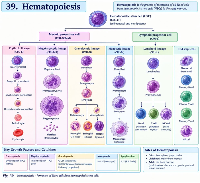
Hematopoiesis is the continuous process by which hematopoietic stem cells produce, differentiate and mature into the various cellular components of blood. In adults, most hematopoiesis occurs in the red bone marrow.
Sites of Hematopoiesis
| Stage of life | Main site | Important feature |
|---|---|---|
| Early embryonic life | Yolk sac | Initial blood-cell formation begins in blood islands. |
| Fetal life | Liver | Liver becomes the major hematopoietic organ. |
| Fetal life | Spleen | Contributes to hematopoiesis, especially during fetal development. |
| Late fetal life and after birth | Bone marrow | Becomes the principal site of hematopoiesis. |
| Adult | Red bone marrow | Predominantly vertebrae, ribs, sternum, pelvis and proximal ends of some long bones. |
Hematopoietic Stem Cell
Hematopoietic stem cells are multipotent cells capable of both self-renewal and differentiation. They give rise to two major progenitor pathways:
Bone Marrow
Bone marrow is a specialized soft tissue present within the medullary cavities of bones and between trabeculae of cancellous bone. Red marrow contains hematopoietic cells supported by a vascular and stromal network. Marrow sinusoids allow mature blood cells to enter the circulation.
Microscopic Features of Red Bone Marrow
- Hematopoietic precursor cells at different stages of maturation.
- Reticular connective-tissue stroma.
- Wide vascular sinusoids lined by endothelial cells.
- Macrophages and stromal cells.
- Megakaryocytes located close to sinusoids.
- Fat cells in variable proportions.
3. Erythropoiesis — Very Important
Formation, maturation and regulation of red blood cells.
Definition
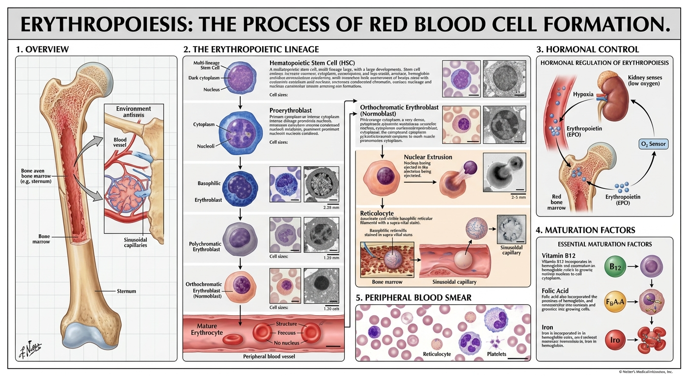
Erythropoiesis is the process of formation and maturation of erythrocytes from hematopoietic stem-cell-derived erythroid progenitors. In adults, it occurs mainly in the red bone marrow.
Stages of Erythropoiesis
| Stage | Main microscopic features | Important change |
|---|---|---|
| Proerythroblast | Large cell, large nucleus, fine chromatin, basophilic cytoplasm. | First recognizable erythroid precursor. |
| Basophilic erythroblast | Smaller cell with strongly basophilic cytoplasm. | Active hemoglobin synthesis begins. |
| Polychromatophilic erythroblast | Cytoplasm becomes gray-blue due to combination of RNA and hemoglobin. | Hemoglobin accumulation increases. |
| Orthochromatic erythroblast | Small cell, condensed dark nucleus, more eosinophilic cytoplasm. | Nucleus is expelled. |
| Reticulocyte | Anucleate young RBC containing residual ribosomal RNA. | Matures in blood within approximately 1–2 days. |
| Mature erythrocyte | Biconcave, anucleate cell with eosinophilic cytoplasm. | Specialized for gas transport. |
Major Changes During Erythrocyte Maturation
- Progressive reduction in cell size.
- Progressive condensation and eventual extrusion of the nucleus.
- Decrease in ribosomal RNA.
- Increasing hemoglobin concentration.
- Change from basophilic to eosinophilic cytoplasm.
- Development of the characteristic biconcave shape.
Regulation by Erythropoietin
Erythropoietin (EPO) is the major hormone regulating erythropoiesis. In adults, it is produced mainly by specialized interstitial cells in the kidney in response to reduced tissue oxygen availability. A smaller contribution comes from the liver.
Requirements for Normal Erythropoiesis
- Iron — essential for hemoglobin synthesis.
- Vitamin B12 — required for normal DNA synthesis.
- Folate — required for DNA synthesis.
- Adequate protein and amino acids.
- Vitamin B6 — involved in heme synthesis.
- Healthy bone marrow.
- Erythropoietin.
- Normal renal function for adequate EPO production.
4. Composition of Blood
Plasma and formed elements.
Definition
Blood is a specialized fluid connective tissue that circulates through the cardiovascular system. It consists of plasma and formed elements.
| Component | Major constituents | Main functions |
|---|---|---|
| Plasma | Water, proteins, electrolytes, nutrients, hormones and waste products. | Transport, osmotic balance, pH regulation and coagulation. |
| RBCs | Erythrocytes containing hemoglobin. | Transport oxygen and contribute to carbon dioxide transport. |
| WBCs | Neutrophils, eosinophils, basophils, lymphocytes and monocytes. | Defense and immune responses. |
| Platelets | Cytoplasmic fragments derived from megakaryocytes. | Primary hemostasis and coagulation. |
5. Red Blood Cells
Structure
Mature RBCs are approximately 7–8 µm in diameter and have a biconcave disc shape. They lack a nucleus, mitochondria and most other organelles. This allows more space for hemoglobin and enables flexibility while passing through narrow capillaries.
Functions
- Transport oxygen from lungs to tissues.
- Transport part of carbon dioxide from tissues to lungs.
- Contribute to blood buffering and acid-base regulation.
- Participate in nitric oxide-related vascular regulation.
Life Span
The average life span of a circulating RBC is approximately 120 days. Senescent RBCs are removed mainly by macrophages in the spleen, liver and bone marrow.
6. White Blood Cells
Leukocytes are nucleated blood cells involved in innate and adaptive defense. They can leave the circulation and enter tissues through processes such as margination, adhesion and diapedesis.
| Cell | Microscopic appearance | Main functions |
|---|---|---|
| Neutrophil | Multilobed nucleus with fine pale granules. | Rapid phagocytosis and killing of bacteria; acute inflammation. |
| Eosinophil | Bilobed nucleus and prominent eosinophilic granules. | Defense against helminths and modulation of allergic inflammation. |
| Basophil | Dark basophilic granules often partially obscure nucleus. | Histamine and mediator release; hypersensitivity reactions. |
| Lymphocyte | Large round nucleus with thin rim of cytoplasm. | Adaptive and innate immune responses; B, T and NK cells. |
| Monocyte | Large cell with kidney-shaped or horseshoe-shaped nucleus. | Precursor of tissue macrophages; phagocytosis and antigen presentation. |
Granulocytes and Agranulocytes
| Granulocytes | Agranulocytes |
|---|---|
| Neutrophils | Lymphocytes |
| Eosinophils | Monocytes |
| Basophils | — |
7. Platelets and Thrombopoiesis
Platelets are small, anucleate cytoplasmic fragments derived from megakaryocytes in the bone marrow. Their major role is maintenance of vascular integrity and primary hemostasis.
Formation
Functions of Platelets
- Adhere to damaged vascular surfaces.
- Become activated after adhesion.
- Release mediators that promote platelet recruitment.
- Form the primary platelet plug.
- Provide a phospholipid surface for coagulation reactions.
- Participate in clot retraction.
- Release growth factors involved in tissue repair.
8. Hemostasis
One of the most important physiology long questions.
Definition
Hemostasis is the physiological process that prevents excessive blood loss after vascular injury while maintaining blood in a fluid state within intact vessels.
Major Stages
1. Vascular Spasm
Immediately after vascular injury, contraction of vascular smooth muscle reduces blood flow. Vasoconstriction is promoted by local neural reflexes, endothelin and mediators released from platelets and damaged tissues.
2. Primary Hemostasis
Primary hemostasis results in formation of a temporary platelet plug.
Platelet Adhesion
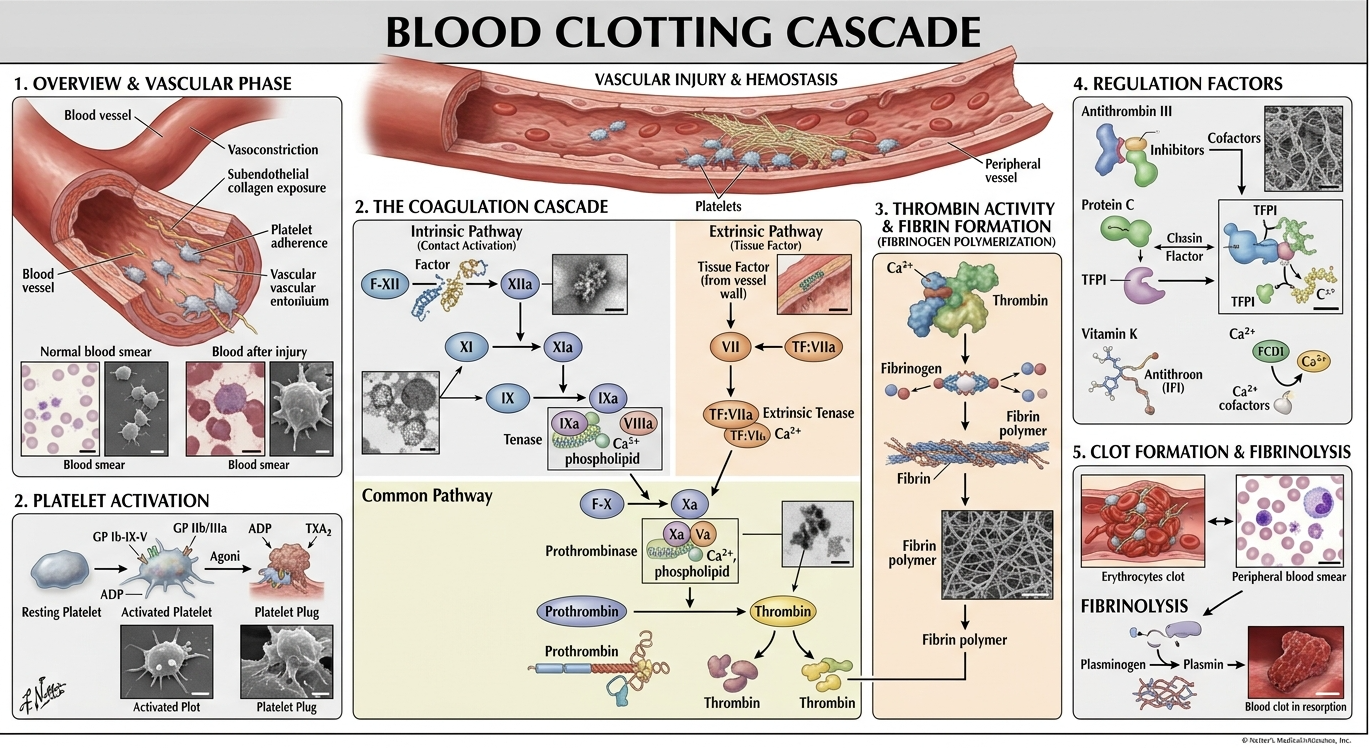
Exposure of subendothelial collagen following endothelial injury promotes platelet adhesion. von Willebrand factor (vWF) helps platelets attach to the injured surface through platelet glycoprotein receptors.
Platelet Activation
Adherent platelets change shape and become activated. They release or generate substances including ADP, thromboxane A₂ and other mediators that promote additional platelet recruitment.
Platelet Aggregation
Activated platelets express conformationally active glycoprotein IIb/IIIa receptors. Fibrinogen can bridge adjacent platelets, producing platelet aggregation and formation of the primary platelet plug.
9. Blood Clotting Mechanism — Very Important 10-Mark Topic
Intrinsic, extrinsic and common pathways with fibrin formation.
Definition of Coagulation
Blood coagulation is a series of enzymatic reactions in which inactive plasma coagulation factors are sequentially activated, ultimately producing thrombin, which converts soluble fibrinogen into insoluble fibrin.
Coagulation Factors
| Factor | Name | Major role |
|---|---|---|
| I | Fibrinogen | Precursor of fibrin. |
| II | Prothrombin | Precursor of thrombin. |
| III | Tissue factor | Initiates extrinsic pathway. |
| IV | Calcium | Required in several coagulation reactions. |
| V | Proaccelerin | Cofactor in prothrombinase complex. |
| VII | Proconvertin | Major factor in extrinsic pathway. |
| VIII | Antihemophilic factor A | Cofactor in intrinsic pathway. |
| IX | Christmas factor | Intrinsic pathway. |
| X | Stuart-Prower factor | Begins common pathway with factor V. |
| XI | Plasma thromboplastin antecedent | Intrinsic pathway. |
| XII | Hageman factor | Contact activation pathway. |
| XIII | Fibrin-stabilizing factor | Cross-links and stabilizes fibrin. |
Extrinsic Pathway
The extrinsic pathway is initiated rapidly following tissue injury. Damaged tissue exposes tissue factor (factor III). Tissue factor combines with activated factor VII in the presence of calcium and phospholipid to activate factor X.
Intrinsic Pathway
The intrinsic pathway involves factors already present in the blood. Contact activation initiates a sequence involving factors XII, XI and IX. Activated factor IX combines with factor VIII, calcium and phospholipid on an appropriate surface to activate factor X.
Common Pathway
Both intrinsic and extrinsic pathways converge at activation of factor X.
Role of Thrombin
- Converts fibrinogen into fibrin.
- Activates factor XIII, promoting fibrin stabilization.
- Activates platelets.
- Amplifies coagulation by activating additional cofactors.
Fibrin Stabilization
Factor XIIIa cross-links fibrin strands and converts the initially unstable fibrin network into a stronger and more stable clot.
Clot Retraction
Platelets contract through their cytoskeletal machinery and pull on the fibrin network. This causes the clot to become smaller and draws the wound margins closer together.
Fibrinolysis
After tissue repair, the clot is gradually removed by fibrinolysis. Plasminogen is converted to plasmin, mainly through tissue plasminogen activator (tPA). Plasmin breaks down fibrin into degradation products.
Natural Anticoagulant Mechanisms
- Antithrombin inhibits thrombin and several activated coagulation factors.
- Protein C and Protein S form an anticoagulant regulatory system.
- Tissue factor pathway inhibitor limits tissue-factor-mediated coagulation.
- Intact endothelium provides antithrombotic and anticoagulant properties.
10. Blood Groups
ABO Blood Group System
The ABO system is determined by the presence or absence of A and B antigens on the RBC membrane and naturally occurring antibodies in plasma.
| Blood group | RBC antigen | Plasma antibody |
|---|---|---|
| A | A | Anti-B |
| B | B | Anti-A |
| AB | A and B | Neither anti-A nor anti-B |
| O | Neither A nor B | Anti-A and anti-B |
Rh Blood Group System
The Rh system contains several antigens, of which the D antigen is the most clinically important. A person with D antigen is generally termed Rh-positive, while absence of D antigen is termed Rh-negative.
Rh Incompatibility
Rh incompatibility may occur when an Rh-negative mother carries an Rh-positive fetus. Fetomaternal hemorrhage can expose the mother to Rh(D) antigen and lead to formation of anti-D antibodies. In a subsequent pregnancy, maternal IgG antibodies may cross the placenta and destroy fetal RBCs, producing hemolytic disease of the fetus and newborn.
Clinical Importance
- Blood grouping is essential before transfusion.
- Cross-matching reduces the risk of incompatible transfusion.
- ABO incompatibility can produce severe hemolytic transfusion reactions.
- Rh typing is important in pregnancy and transfusion medicine.
11. Lymph
Definition
Lymph is a clear to pale fluid formed from interstitial fluid that enters lymphatic capillaries. It eventually returns to the venous circulation.
Formation of Lymph
Blood pressure at systemic capillaries causes a portion of plasma fluid to move into the interstitial space. Most fluid is returned directly to blood capillaries, while excess interstitial fluid and proteins enter lymphatic capillaries and become lymph.
Functions of Lymph
- Returns excess tissue fluid to the blood.
- Returns leaked plasma proteins to circulation.
- Transports absorbed dietary lipids from intestinal lacteals.
- Provides a pathway for lymphocyte circulation.
- Transports antigens to lymphoid organs for immune surveillance.
- Participates in inflammatory and immune responses.
12. Lymphatic Vessels
Lymphatic Capillaries
Lymphatic capillaries are blind-ended microscopic vessels present in most tissues. Their endothelial cells overlap loosely, creating flap-like openings through which interstitial fluid, proteins and cells can enter.
Collecting Lymphatic Vessels
Larger lymphatic vessels possess thin walls with endothelial lining, connective tissue, smooth muscle and valves. Valves help maintain one-directional flow toward the venous system.
Lymphatic Trunks
Collecting lymphatics converge into larger trunks including jugular, subclavian, bronchomediastinal, intestinal and lumbar trunks.
Major Lymphatic Ducts
| Duct | Major drainage | Termination |
|---|---|---|
| Thoracic duct | Most of the body below the diaphragm and left upper body. | Left venous angle. |
| Right lymphatic duct | Right upper quadrant of the body. | Right venous angle. |
13. Lymph Node — Gross & Microscopic Anatomy
Gross Anatomy
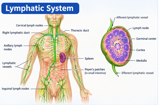
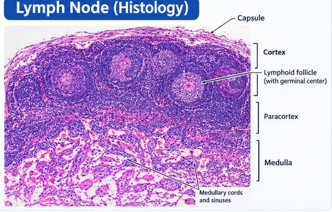
A lymph node is a small, bean-shaped lymphoid organ located along lymphatic vessels. It receives lymph through afferent lymphatic vessels and drains lymph through efferent lymphatic vessels at the hilum.
Microscopic Structure
1. Capsule
The lymph node is surrounded by a connective-tissue capsule. Trabeculae extend inward from the capsule and provide structural support.
2. Cortex
The outer cortex contains lymphoid follicles. Primary follicles contain mainly small lymphocytes, while secondary follicles possess germinal centers containing activated and proliferating B lymphocytes.
3. Paracortex
The paracortex lies between cortex and medulla and is particularly rich in T lymphocytes. High endothelial venules provide an important route through which circulating lymphocytes enter lymph nodes.
4. Medulla
The medulla contains medullary cords and medullary sinuses. Medullary cords contain lymphocytes, plasma cells and macrophages, while sinuses allow lymph to flow toward the efferent lymphatic vessel.
Lymph Flow Through a Node
Functions
- Filters lymph.
- Traps foreign particles and microorganisms.
- Provides a site for antigen presentation.
- Supports B-cell and T-cell activation.
- Supports antibody production through plasma cells.
14. Spleen — Gross & Microscopic Anatomy
Gross Anatomy
The spleen is the largest lymphoid organ. It is located in the left upper quadrant of the abdomen, beneath the diaphragm and close to the stomach. It has diaphragmatic and visceral surfaces and a hilum through which vessels enter and leave.
Microscopic Structure
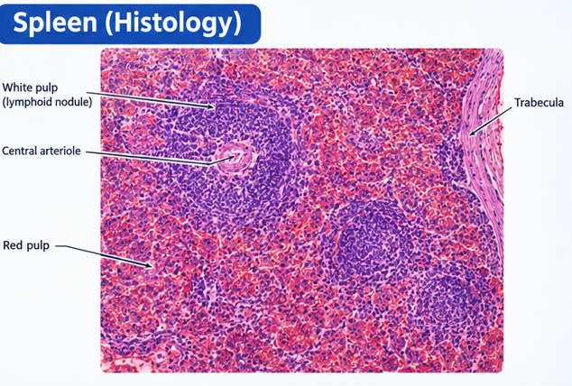
The spleen is surrounded by a connective-tissue capsule from which trabeculae extend into the organ. Its functional tissue is called splenic pulp, divided into white pulp and red pulp.
White Pulp
White pulp consists of lymphoid tissue surrounding branches of the splenic arterial circulation. The periarteriolar lymphoid sheath (PALS) is particularly rich in T lymphocytes. Lymphoid follicles contain predominantly B lymphocytes.
Marginal Zone
The marginal zone surrounds the white pulp and contains macrophages, lymphocytes and antigen-presenting cells. It serves as an important interface between circulating blood and lymphoid tissue.
Red Pulp
Red pulp consists of splenic cords and venous sinusoids. It filters circulating blood and removes old, damaged or abnormal erythrocytes. Macrophages in the red pulp phagocytose senescent RBCs and help recycle their components.
Functions of Spleen
- Filters blood.
- Removes senescent and damaged RBCs.
- Participates in immune responses to blood-borne antigens.
- Contains large populations of lymphocytes and macrophages.
- Recycles iron from hemoglobin.
- Acts as a reservoir for some blood components.
15. Thymus — Gross & Microscopic Anatomy
Gross Anatomy
The thymus is a primary lymphoid organ located in the anterior superior mediastinum behind the sternum. It is relatively large during childhood and gradually undergoes involution after puberty, with increasing replacement by adipose tissue.
Microscopic Anatomy
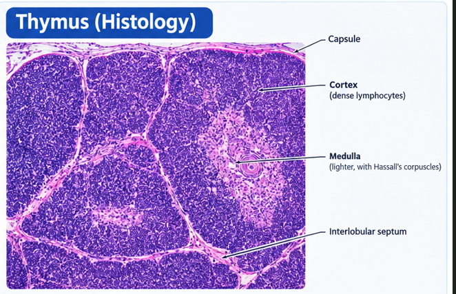
A connective-tissue capsule surrounds the thymus and sends septa into the organ, dividing it into lobules.
Cortex
The cortex is densely packed with immature T lymphocytes (thymocytes). It contains epithelial reticular cells that form an important structural and functional environment for T-cell development.
Medulla
The medulla contains fewer lymphocytes and more epithelial reticular cells. Characteristic Hassall's corpuscles are found in the thymic medulla.
Functions
- Site of T-lymphocyte maturation.
- Provides an environment for T-cell selection.
- Helps establish central immune tolerance.
- Produces thymic factors involved in lymphocyte development.
16. Tonsils
Tonsils are aggregates of lymphoid tissue positioned around the entrance to the respiratory and digestive tracts. They contribute to immune surveillance of inhaled and ingested antigens.
| Tonsil | Location | Important microscopic feature |
|---|---|---|
| Palatine tonsils | Lateral wall of oropharynx | Stratified squamous non-keratinized epithelium and deep crypts. |
| Pharyngeal tonsil | Roof/posterior wall of nasopharynx | Respiratory epithelium with lymphoid tissue. |
| Lingual tonsils | Posterior tongue | Lymphoid follicles with associated mucosal glands. |
Functions
- Sample antigens entering through mouth and nose.
- Support local lymphocyte activation.
- Participate in mucosal immune defense.
17. Bone Marrow — Gross & Microscopic Anatomy
Types
- Red marrow: actively produces blood cells.
- Yellow marrow: contains a greater proportion of adipose tissue.
Microscopic Features
- Hematopoietic cell cords between vascular sinusoids.
- Reticular stromal framework.
- Macrophages.
- Megakaryocytes adjacent to marrow sinusoids.
- Developing erythroid and myeloid cells.
- Adipocytes.
Functions
- Produces RBCs.
- Produces WBCs.
- Produces platelets through megakaryocytes.
- Provides an important microenvironment for hematopoietic stem cells.
18. Reticuloendothelial System / Mononuclear Phagocyte System
Definition
The term reticuloendothelial system historically described cells with phagocytic properties distributed throughout the body. The more precise modern term is the mononuclear phagocyte system (MPS), which includes circulating monocytes and tissue macrophages.
| Cell | Main location | Major function |
|---|---|---|
| Monocyte | Blood | Circulating precursor and phagocytic cell. |
| Macrophage | Many tissues | Phagocytosis, antigen presentation and cytokine production. |
| Kupffer cell | Liver sinusoids | Phagocytosis of blood-borne particles and old RBC components. |
| Splenic macrophage | Spleen | Removal of senescent RBCs and immune surveillance. |
| Lymph-node macrophage | Lymph nodes | Filters lymph and phagocytoses foreign material. |
| Alveolar macrophage | Lung alveoli | Removes inhaled particles and microorganisms. |
| Microglia | Central nervous system | Resident phagocytic immune cells of CNS. |
Functions of MPS
- Phagocytosis of microorganisms.
- Removal of dead and damaged cells.
- Removal of senescent RBCs.
- Antigen processing and presentation.
- Cytokine production.
- Participation in inflammation and tissue repair.
19. Immune System
Organization of Immune Organs
| Category | Organs/tissues | Main role |
|---|---|---|
| Primary lymphoid organs | Bone marrow and thymus | Development and maturation of lymphocytes. |
| Secondary lymphoid organs | Lymph nodes, spleen, tonsils and other lymphoid tissues | Antigen encounter and activation of mature lymphocytes. |
Innate Immunity
Innate immunity provides rapid, non-specific defense. Important components include epithelial barriers, phagocytes, natural killer cells, complement proteins, inflammatory mediators and pattern-recognition receptors.
Adaptive Immunity
Adaptive immunity is antigen-specific and develops immunological memory. It includes humoral immunity mediated mainly by B lymphocytes and antibodies, and cell-mediated immunity mediated mainly by T lymphocytes.
B Lymphocytes
B cells can differentiate into plasma cells that secrete antibodies. They also contribute to immunological memory and antigen presentation.
T Lymphocytes
- Helper T cells: coordinate immune responses through cytokines.
- Cytotoxic T cells: kill infected or abnormal cells.
- Regulatory T cells: help control excessive immune responses.
- Memory T cells: provide long-term immunological memory.
20. Important 10-Mark Long Questions
Answer-ready MSc/TU-style preparation.
Blood consists of plasma and formed elements. Plasma contains water, proteins, electrolytes, nutrients, hormones and metabolic products. Formed elements include RBCs, WBCs and platelets.
| Cell | Important structural feature | Major function |
|---|---|---|
| RBC | Biconcave, anucleate cell containing hemoglobin. | O₂ and CO₂ transport. |
| Neutrophil | Multilobed nucleus. | Phagocytosis and acute inflammation. |
| Eosinophil | Bilobed nucleus and eosinophilic granules. | Parasite defense and allergy modulation. |
| Basophil | Dark cytoplasmic granules. | Inflammatory and hypersensitivity reactions. |
| Lymphocyte | Large nucleus with scant cytoplasm. | Adaptive and innate immunity. |
| Monocyte | Large cell with kidney-shaped nucleus. | Phagocytosis and macrophage precursor. |
| Platelet | Anucleate cytoplasmic fragment. | Hemostasis and clot formation. |
Hemostasis occurs through vascular constriction, platelet plug formation, coagulation, clot retraction and fibrinolysis.
Primary Hemostasis
Vascular injury exposes subendothelial structures. Platelets adhere with assistance from vWF, become activated and release mediators. Activated platelets recruit additional platelets and aggregate to form a temporary platelet plug.
Secondary Hemostasis
Coagulation reactions generate thrombin. Thrombin converts fibrinogen into fibrin. Factor XIII stabilizes the fibrin network.
Pathways
The extrinsic pathway is initiated by tissue factor and factor VII. The intrinsic pathway involves factors XII, XI, IX and VIII. Both pathways converge at factor X and enter the common pathway.
After tissue repair, plasmin-mediated fibrinolysis removes the fibrin clot. Natural anticoagulant mechanisms prevent uncontrolled coagulation.
Extrinsic Pathway
Tissue injury exposes tissue factor. Tissue factor combines with factor VII and calcium and activates factor X.
Intrinsic Pathway
Contact activation initiates factor XII. The sequence proceeds through XI and IX. Activated IX combines with VIII, calcium and phospholipid to activate factor X.
Common Pathway
Factor Xa combines with factor Va, calcium and phospholipid to form the prothrombinase complex. This converts prothrombin to thrombin. Thrombin converts fibrinogen into fibrin, and factor XIII stabilizes fibrin.
The lymphatic system is a network of capillaries, vessels, trunks, ducts and lymphoid organs. It maintains fluid balance, transports absorbed lipids and participates in immune defense.
Lymphatic Capillaries
These are blind-ended vessels with specialized endothelial junctions allowing entry of interstitial fluid and proteins.
Collecting Vessels
Larger vessels contain valves and smooth muscle and transport lymph toward regional lymph nodes and lymphatic trunks.
Major Ducts
The thoracic duct drains most of the body and enters the left venous angle. The right lymphatic duct drains the right upper quadrant and enters the right venous angle.
Functions
- Fluid balance.
- Protein return.
- Lipid absorption.
- Immune-cell circulation.
- Transport of antigens.
Lymph nodes are encapsulated lymphoid organs positioned along lymphatic vessels. They receive lymph through afferent vessels and drain it through efferent vessels.
Microscopic Structure
- Connective-tissue capsule.
- Trabeculae.
- Outer cortex containing lymphoid follicles.
- Paracortex rich in T lymphocytes.
- Medullary cords containing lymphocytes, plasma cells and macrophages.
- Medullary sinuses.
- Afferent and efferent lymphatic vessels.
Functions
Lymph nodes filter lymph, trap antigens and microorganisms, provide sites for antigen presentation and support activation of B and T lymphocytes.
The spleen is the largest lymphoid organ and lies in the left upper quadrant of the abdomen.
Microscopic Structure
The organ has a capsule and trabeculae. Its parenchyma is divided into white pulp and red pulp. White pulp contains lymphoid tissue around arterial branches, including PALS and lymphoid follicles. Red pulp consists of splenic cords and venous sinusoids.
Functions
- Blood filtration.
- Removal of senescent RBCs.
- Immune responses to blood-borne antigens.
- Macrophage-mediated phagocytosis.
- Iron recycling.
The thymus is a primary lymphoid organ in the anterior superior mediastinum. It is divided into lobules by connective-tissue septa.
The cortex is densely populated by immature T lymphocytes, while the medulla contains fewer lymphocytes and characteristic Hassall's corpuscles. Epithelial reticular cells support thymocyte development.
The major function of thymus is maturation and selection of T lymphocytes, contributing to establishment of central immune tolerance.
The ABO system depends on A and B antigens on RBCs and corresponding naturally occurring antibodies in plasma. Group A has A antigen and anti-B antibody; group B has B antigen and anti-A; group AB has both antigens and neither antibody; group O lacks A and B antigens but has both antibodies.
In the Rh system, the D antigen is clinically important. Rh-positive individuals possess D antigen, whereas Rh-negative individuals lack it.
Incompatible transfusion can cause antigen-antibody reactions and hemolysis. Rh incompatibility can also cause hemolytic disease of the fetus and newborn.
The mononuclear phagocyte system consists mainly of circulating monocytes and tissue macrophages. These cells originate from hematopoietic progenitors and become specialized according to their tissue environment.
| Location | Macrophage | Major function |
|---|---|---|
| Liver | Kupffer cells | Remove blood-borne particles and damaged RBC components. |
| Spleen | Splenic macrophages | Remove senescent RBCs and monitor blood antigens. |
| Lymph node | Sinus macrophages | Filter lymph. |
| Lung | Alveolar macrophages | Remove inhaled particles and microorganisms. |
| CNS | Microglia | Resident immune surveillance and phagocytosis. |
| Primary organs | Function |
|---|---|
| Bone marrow | Hematopoiesis and B-cell development. |
| Thymus | T-cell maturation and selection. |
| Secondary organs | Function |
|---|---|
| Lymph nodes | Filter lymph and initiate immune responses. |
| Spleen | Filters blood and responds to blood-borne antigens. |
| Tonsils | Monitor antigens entering through respiratory and digestive pathways. |
21. Important 5-Mark Questions
Erythropoiesis
Define erythropoiesis, list stages and explain the role of EPO.
Hematopoiesis
Describe sites of hematopoiesis during fetal and adult life.
RBC
Describe structure and functions of erythrocytes.
WBC
Classify WBCs and mention their major functions.
Platelets
Describe formation and functions of platelets.
Hemostasis
Describe the major stages of hemostasis.
Coagulation
Explain intrinsic and extrinsic pathways briefly.
ABO system
Explain ABO blood groups with antigen and antibody distribution.
Rh system
Explain Rh blood group and Rh incompatibility.
Lymph
Define lymph and discuss its composition and functions.
Lymphatic vessels
Describe lymphatic capillaries, vessels and major ducts.
Lymph node
Describe cortex, paracortex and medulla.
Spleen
Explain white pulp and red pulp.
Thymus
Describe cortex, medulla and Hassall's corpuscles.
MPS
Describe the mononuclear phagocyte system and examples.
Immune organs
Differentiate primary and secondary lymphoid organs.
22. Important 2-Mark Questions
Blood
- Define blood.
- Name the formed elements of blood.
- What is plasma?
- What is hematopoiesis?
- Name the major sites of adult hematopoiesis.
- What is erythropoietin?
- What is a reticulocyte?
- What is the life span of an RBC?
Blood Cells
- State two functions of RBCs.
- Name the granulocytes.
- Name the agranulocytes.
- What is a platelet?
- State two functions of neutrophils.
- State one function of eosinophils.
- What are monocytes?
Hemostasis
- Define hemostasis.
- What is platelet adhesion?
- What is platelet aggregation?
- What is tissue factor?
- What is thrombin?
- What is fibrin?
- What is fibrinolysis?
- What is factor XIII?
Blood Groups
- What is an antigen?
- What is an antibody?
- Name the four ABO blood groups.
- Which antigen is important in Rh typing?
- What is Rh incompatibility?
- What is cross-matching?
Lymphatic System
- Define lymph.
- State two functions of lymph.
- What is a lymphatic capillary?
- Name the two major lymphatic ducts.
- What is the thoracic duct?
- What is a lymph node?
Lymphoid Organs
- Name the primary lymphoid organs.
- Name three secondary lymphoid organs.
- What is white pulp?
- What is red pulp?
- What are Hassall's corpuscles?
- What is PALS?
23. High-Yield Microscopic Anatomy Comparison
| Organ | Key microscopic structures | Major function |
|---|---|---|
| Lymph node | Capsule, cortex, follicles, paracortex, medullary cords and sinuses. | Filters lymph and supports lymphocyte activation. |
| Spleen | Capsule, trabeculae, white pulp, marginal zone, red pulp, sinusoids. | Filters blood and removes old RBCs. |
| Thymus | Lobules, cortex, medulla, epithelial reticular cells and Hassall's corpuscles. | T-cell maturation. |
| Bone marrow | Hematopoietic cells, sinusoids, stromal cells and megakaryocytes. | Blood-cell production. |
| Tonsil | Lymphoid follicles, surface epithelium and, depending on type, crypts. | Mucosal immune surveillance. |
24. Blood vs Lymph
| Feature | Blood | Lymph |
|---|---|---|
| Color | Red due to hemoglobin-containing RBCs. | Usually clear or pale. |
| Main location | Blood vessels. | Lymphatic vessels. |
| RBCs | Normally abundant. | Normally absent in ordinary lymph. |
| Major function | Transport and homeostasis. | Fluid return and immune surveillance. |
| Direction | Closed cardiovascular circulation. | One-way return toward venous circulation. |
25. Must-Memorize Flow Diagrams
Erythropoiesis
Hemostasis
Common Coagulation Pathway
Lymph Flow
26. Important Exam Points — Final Revision
⭐ Erythropoiesis
Remember all stages, morphological changes and EPO regulation.
⭐ Coagulation
Know intrinsic, extrinsic and common pathways and major coagulation factors.
⭐ Hemostasis
Know vasoconstriction → platelet plug → coagulation → fibrin clot → fibrinolysis.
⭐ Lymph Node
Cortex, paracortex, medulla, follicles, sinuses and functions are essential for histology questions.
⭐ Spleen
White pulp, PALS, marginal zone, red pulp, splenic cords and sinusoids.
⭐ Thymus
Cortex, medulla, epithelial reticular cells and Hassall's corpuscles.
⭐ Blood Groups
ABO antigens/antibodies, Rh D antigen, transfusion compatibility and Rh incompatibility.
⭐ MPS
Monocytes, macrophages and tissue-specific macrophages such as Kupffer cells.
- Erythropoiesis and its regulation.
- Hemostasis and blood coagulation mechanism.
- Intrinsic, extrinsic and common coagulation pathways.
- Lymphatic system and lymphatic circulation.
- Gross and microscopic anatomy of lymph node.
- Gross and microscopic anatomy of spleen.
- Gross and microscopic anatomy of thymus.
- ABO and Rh blood groups.
- Composition and functions of blood cells.
- Reticuloendothelial/mononuclear phagocyte system.
27. Important Figures to Prepare
Blood & Hematology
- Hematopoiesis diagram
- Erythropoiesis stages
- Blood-cell morphology
- Platelet formation
- Coagulation cascade
Lymphatic System
- Complete lymphatic system
- Lymphatic capillary
- Thoracic duct
- Lymph-node structure
- Lymph flow through lymph node
Lymphoid Organs
- Spleen histology
- Thymus histology
- Tonsil histology
- Bone marrow histology
Immune System
- Primary and secondary lymphoid organs
- B-cell pathway
- T-cell pathway
- Mononuclear phagocyte system