Complete MSc Medical Microbiology examination notes covering
gross and microscopic anatomy, respiratory mechanics, pulmonary
ventilation, gas exchange, partial pressures, oxygen and carbon
dioxide transport, circulation and regulation of breathing.
Why Respiratory System Is an Important LQ Topic
MB 551 syllabus:
Different parts of respiratory system; gross and microscopic
anatomy of different parts; functions of parts.
For examination preparation, these anatomical topics should be
connected with the major physiological processes of respiration,
particularly pulmonary ventilation, mechanics of breathing,
gas exchange and transport of respiratory gases.
Very important 10-mark areas:
Gross and microscopic anatomy of respiratory system.
Structure and functions of nose, pharynx, larynx, trachea and bronchi.
Gross and microscopic anatomy of lungs and alveoli.
Mechanism of breathing.
Pulmonary ventilation and alveolar ventilation.
Lung volumes and capacities.
Gas exchange between alveoli and blood.
Partial pressure of oxygen and carbon dioxide.
Transport of oxygen in blood.
Transport of carbon dioxide in blood.
Oxygen-haemoglobin dissociation curve and Bohr effect.
The respiratory system is an organ system responsible primarily for
exchange of respiratory gases between the atmosphere and blood.
It supplies oxygen required for cellular metabolism and removes
carbon dioxide produced during cellular respiration.
Respiration is not limited to movement of air into and out of the lungs.
It involves several coordinated processes including pulmonary
ventilation, diffusion of gases between alveoli and blood, transport
of oxygen and carbon dioxide by blood, and exchange of gases between
systemic blood and tissues.
Major Processes of Respiration
Pulmonary ventilation:
movement of air into and out of lungs.
External respiration:
exchange of O₂ and CO₂ between alveoli and pulmonary blood.
Gas transport:
transport of O₂ and CO₂ through blood.
Internal respiration:
exchange of gases between systemic blood and tissues.
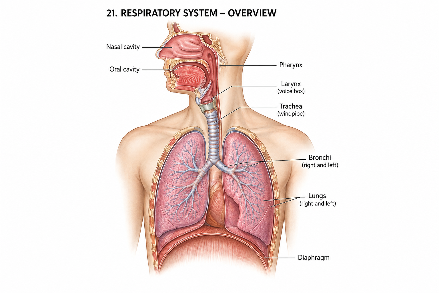
Figure: Realistic labelled overview of the human respiratory system.
2. Different Parts of Respiratory System
Anatomically, the respiratory system can be divided into the upper
and lower respiratory tracts.
Division
Parts
Main role
Upper respiratory tract
Nose, nasal cavity, paranasal sinuses and pharynx
Air entry, filtration, warming, humidification and conduction
Lower respiratory tract
Larynx, trachea, bronchi, bronchioles and lungs
Air conduction and gas exchange
Functional Division
Conducting Zone
The conducting zone extends from the nose to the terminal bronchioles.
It conducts air but normally does not participate directly in gas
exchange.
Its major functions include filtration, warming, humidification and
distribution of inspired air.
Respiratory Zone
The respiratory zone includes respiratory bronchioles, alveolar ducts,
alveolar sacs and alveoli. This is the principal region where
gas exchange occurs.
3. Nose and Nasal Cavity
Gross Anatomy
The nose forms the principal entrance for air into the respiratory
tract. The nasal cavity is divided into right and left halves by
the nasal septum.
Each nasal cavity contains three major conchae:
superior, middle and inferior nasal conchae. These structures increase
the surface area and create turbulence, helping contact inspired air
with the mucosa.
Microscopic Anatomy
Most of the respiratory region is lined by pseudostratified ciliated
columnar epithelium containing goblet cells.
Ciliated columnar cells move mucus toward the pharynx.
Goblet cells secrete mucus.
Basal cells act as progenitor cells.
Numerous blood vessels help warm inspired air.
Functions
Provides passage for inspired air.
Filters particles.
Warms air.
Humidifies air.
Contributes to olfaction.
Provides resonance for speech.
4. Pharynx
The pharynx is a muscular tube that serves as a common passage for
respiratory and digestive pathways.
Parts
Nasopharynx
Oropharynx
Laryngopharynx
Functions
Conducts air from nasal cavity toward larynx.
Participates in swallowing.
Contributes to resonance of voice.
Epithelium
The nasopharynx is primarily lined by respiratory epithelium,
while regions exposed to greater mechanical stress, particularly
the oropharynx and laryngopharynx, are lined predominantly by
stratified squamous epithelium.
5. Larynx
The larynx is a cartilaginous organ situated between the pharynx
and trachea. It maintains an open airway and plays an important
role in phonation.
Major Cartilages
Thyroid cartilage
Cricoid cartilage
Epiglottis
Arytenoid cartilages
Vocal Cords
The larynx contains vestibular folds and vocal folds. The true vocal
folds contain skeletal muscle and elastic tissue and are involved
in sound production.
Microscopic Anatomy
Much of the laryngeal lining contains respiratory epithelium.
However, areas exposed to significant mechanical stress, especially
the true vocal folds, are covered by stratified squamous epithelium.
Functions
Maintains airway.
Produces voice.
Protects lower respiratory tract during swallowing.
Participates in cough reflex.
6. Trachea
Gross Anatomy
The trachea is a flexible air-conducting tube extending from the
larynx to the main bronchi. It is supported by a series of
incomplete C-shaped cartilage rings.
The posterior portion of each cartilage ring is completed by
trachealis smooth muscle and connective tissue.
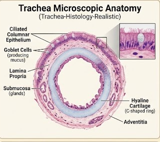
Microscopic Anatomy
The tracheal wall contains several layers.
Mucosa:
pseudostratified ciliated columnar epithelium with goblet cells.
Figure: Realistic histological structure of the trachea.
7. Bronchi and Bronchioles
Bronchi
The trachea divides into right and left main bronchi. The right main
bronchus is generally shorter, wider and more vertically oriented
than the left.
The main bronchi divide repeatedly into lobar and segmental bronchi,
which continue into smaller bronchi and bronchioles.
Microscopic Features of Bronchi
Respiratory epithelium.
Goblet cells.
Smooth muscle.
Seromucous glands.
Cartilage plates.
Bronchioles
Bronchioles are smaller airways that lack cartilage and submucosal
glands. Their epithelium becomes progressively shorter as the airway
diameter decreases.
Smooth muscle becomes relatively important in bronchioles and helps
regulate airway diameter.
Terminal Bronchioles
Terminal bronchioles represent the final part of the conducting zone.
They are lined mainly by simple cuboidal epithelium and contain
club cells.
Functions
Conduct air deeper into the lungs.
Distribute air to different lung regions.
Regulate airflow through smooth muscle.
8. Gross Anatomy of the Lungs
The lungs are paired respiratory organs situated within the thoracic
cavity. They are separated by the mediastinum and covered by pleura.
Right Lung
The right lung has three lobes:
superior, middle and inferior. These are separated by horizontal
and oblique fissures.
Left Lung
The left lung has two lobes:
superior and inferior. It contains the cardiac notch and lingula.
The left lung is smaller than the right because of the position
of the heart.
Surfaces
Costal surface
Mediastinal surface
Diaphragmatic surface
Hilum and Root
The hilum is the region on the mediastinal surface through which
bronchi, pulmonary vessels, lymphatics and nerves enter or leave
the lung.
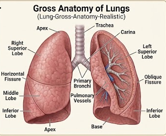
Figure: Gross anatomy of right and left lungs.
9. Microscopic Anatomy of Respiratory Tract
General Change in Epithelium
As the conducting airway becomes progressively smaller, the
respiratory epithelium becomes shorter and simpler.
Region
Predominant epithelium
Important feature
Trachea
Pseudostratified ciliated columnar
Goblet cells, cartilage, glands
Bronchi
Pseudostratified ciliated columnar
Cartilage plates and smooth muscle
Bronchioles
Simple columnar/cuboidal
No cartilage or submucosal glands
Terminal bronchioles
Simple cuboidal
Club cells
Respiratory bronchioles
Low cuboidal
Alveoli begin appearing
Alveoli
Simple squamous epithelium
Specialized for gas exchange
10. Alveoli and Respiratory Membrane
Alveoli are microscopic air sacs forming the principal site of
gas exchange in the lungs. They provide a very large surface area
and have extremely thin walls.
Types of Alveolar Cells
Type I Pneumocytes
Type I pneumocytes are thin squamous cells covering most of the
alveolar surface. They form the major diffusion barrier for
respiratory gases.
Type II Pneumocytes
Type II pneumocytes are more cuboidal cells that produce pulmonary
surfactant. Surfactant reduces surface tension and helps prevent
alveolar collapse.
Alveolar Macrophages
Alveolar macrophages remove inhaled particles and microorganisms
and help maintain cleanliness of the alveolar spaces.
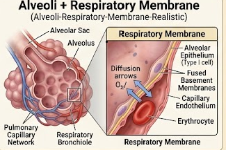
Figure: Alveolus and respiratory membrane showing the relationship
between alveolar air and pulmonary capillary blood.
Respiratory Membrane
The respiratory membrane is the thin barrier across which oxygen and
carbon dioxide diffuse between alveolar air and pulmonary capillary
blood.
It consists functionally of the alveolar epithelium, fused basement
membrane/interstitial components and capillary endothelium.
Factors favoring gas diffusion:
Large surface area.
Very thin respiratory membrane.
Large partial-pressure gradient.
Good pulmonary perfusion.
Appropriate ventilation of alveoli.
11. Functions of Different Parts of Respiratory System
Part
Major functions
Nose and nasal cavity
Air entry, filtration, warming, humidification and olfaction.
Pharynx
Conducts air and contributes to swallowing and resonance.
Larynx
Maintains airway, protects lower respiratory tract and produces voice.
Trachea
Conducts air and provides mucociliary clearance.
Bronchi
Distribute air to different lung regions.
Bronchioles
Regulate airway resistance and distribute air toward respiratory regions.
Alveoli
Main site of oxygen and carbon dioxide exchange.
Lungs
Provide the organ surface and vascular network necessary for gas exchange.
Pleura
Reduces friction and contributes to mechanical coupling of lungs to thoracic wall.
12. Pulmonary Ventilation
Pulmonary ventilation is the movement of air between the atmosphere
and alveoli. It consists of two phases:
inspiration and expiration.
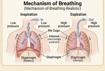
Inspiration
During normal inspiration, the diaphragm contracts and moves downward.
The external intercostal muscles contract and elevate the ribs.
Expansion of the thoracic cavity increases lung volume.
According to the pressure-volume relationship, an increase in lung
volume causes a decrease in intrapulmonary pressure. When alveolar
pressure falls below atmospheric pressure, air enters the lungs.
Expiration
Quiet expiration is mainly passive. The diaphragm and external
intercostal muscles relax, and elastic recoil of the lungs decreases
thoracic and lung volume.
Alveolar pressure then becomes slightly greater than atmospheric
pressure and air flows outward.
Forced Breathing
During forced inspiration, accessory inspiratory muscles assist
expansion of the thorax. During forced expiration, internal
intercostal muscles and abdominal muscles increase thoracic pressure
and actively drive air out.
The lungs are separated from the thoracic wall by the pleural cavity.
The intrapleural pressure is normally negative relative to atmospheric
pressure. This negative pressure helps keep the lungs expanded.
Important physiological concept:
Breathing occurs because changes in thoracic volume produce changes
in pressure, and air moves down a pressure gradient.
14. Lung Volumes and Capacities
Lung volumes describe the amount of air moved or remaining within
the lungs during different phases of breathing.
Volume / Capacity
Definition
Approximate adult value
Tidal volume (TV)
Air inspired or expired during normal quiet breathing
~500 mL
Inspiratory reserve volume (IRV)
Additional air that can be inspired after normal inspiration
~3000 mL
Expiratory reserve volume (ERV)
Additional air that can be expired after normal expiration
~1100 mL
Residual volume (RV)
Air remaining after maximal expiration
~1200 mL
Capacities
Capacity
Formula
Approximate value
Inspiratory capacity
TV + IRV
~3500 mL
Functional residual capacity
ERV + RV
~2300 mL
Vital capacity
IRV + TV + ERV
~4600 mL
Total lung capacity
VC + RV
~5800 mL
Exam note:
Exact lung volumes vary with age, sex, height, body size and
physiological condition. The values above are approximate reference
values useful for examination.
15. Alveolar Ventilation
Not all inspired air reaches the gas-exchanging regions of the lungs.
Some remains in the conducting airways and forms the anatomical
dead space.
Minute Ventilation
Minute ventilation is the total volume of air entering or leaving
the respiratory tract per minute.
Alveolar ventilation is more important than minute ventilation
for effective gas exchange because only air reaching functioning
alveoli participates directly in exchange.
16. Pulmonary Circulation and Perfusion
Pulmonary circulation carries deoxygenated blood from the right
ventricle to the lungs and returns oxygenated blood to the left atrium.
Right Ventricle
→
Pulmonary Trunk
→
Pulmonary Arteries
→
Pulmonary Capillaries
→
Pulmonary Veins
→
Left Atrium
Pulmonary capillary blood flows around alveoli and provides the
vascular surface required for gas exchange.
Perfusion
Perfusion refers to blood flow through pulmonary capillaries.
Effective gas exchange requires appropriate matching between
ventilation of alveoli and perfusion of pulmonary capillaries.
17. Gas Exchange in Detail
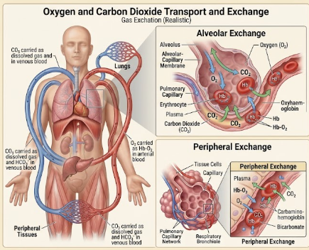
Gas exchange is the movement of oxygen and carbon dioxide across
the respiratory membrane according to their partial-pressure
gradients.
External Respiration
External respiration occurs between alveolar air and pulmonary
capillary blood.
Oxygen moves from alveolar air into blood because the partial pressure
of oxygen is higher in alveolar air than in incoming venous blood.
Carbon dioxide moves from pulmonary capillary blood into alveolar air
because the partial pressure of carbon dioxide is higher in venous
blood than in alveolar air.
Internal Respiration
Internal respiration occurs between systemic capillary blood and
tissue cells. Oxygen moves from blood to tissues, while carbon dioxide
moves from tissues into blood.
Figure: Gas exchange across the alveolar-capillary respiratory membrane.
Factors Affecting Gas Exchange
Partial-pressure gradient.
Thickness of respiratory membrane.
Surface area available for diffusion.
Diffusion coefficient of the gas.
Ventilation-perfusion matching.
Solubility of the gas.
Fick's Law of Diffusion
The rate of diffusion across a biological membrane increases with
surface area and partial-pressure gradient and decreases with
membrane thickness.
Rate of diffusion ∝ (Surface area × Partial-pressure gradient × Diffusion coefficient) / Membrane thickness
18. Partial Pressure of O₂ and CO₂
Partial pressure is the pressure contributed by an individual gas
within a mixture of gases. Respiratory gas exchange occurs because
gases move down their partial-pressure gradients.
Alveolar and Blood Values
Location
PO₂
PCO₂
Atmospheric dry air at sea level
~159 mmHg
~0.3 mmHg
Alveolar air
~100 mmHg
~40 mmHg
Incoming pulmonary arterial blood
~40 mmHg
~45 mmHg
Systemic arterial blood
~95–100 mmHg
~40 mmHg
Typical systemic venous blood
~40 mmHg
~45 mmHg
Why Is Alveolar PO₂ Lower Than Atmospheric PO₂?
Atmospheric air is not identical to alveolar air. Inspired air
becomes humidified in the respiratory tract, and oxygen is continuously
removed from alveolar air into pulmonary blood while carbon dioxide
is continuously added from blood.
Why Does CO₂ Diffuse Despite a Smaller Gradient?
Carbon dioxide is considerably more soluble in biological membranes
than oxygen. Therefore, it diffuses efficiently even with a smaller
partial-pressure gradient.
Direction of diffusion:
O₂: Alveoli → pulmonary blood
CO₂: Pulmonary blood → alveoli
19. Transport of Oxygen in Blood
Oxygen is transported from lungs to tissues mainly in two forms:
dissolved in plasma and reversibly bound to haemoglobin in red blood cells.
1. Dissolved Oxygen
A small proportion of oxygen is physically dissolved in plasma.
Although quantitatively small, dissolved oxygen determines the
measured partial pressure of oxygen in blood.
2. Haemoglobin-Bound Oxygen
Most oxygen is transported bound reversibly to the iron-containing
haem groups of haemoglobin.
Each haemoglobin molecule can bind up to four oxygen molecules.
Hb + O₂ ⇌ HbO₂
Oxygen Loading in Lungs
In pulmonary capillaries, the relatively high PO₂ promotes binding
of oxygen to haemoglobin.
Oxygen Unloading in Tissues
In systemic tissues, the lower PO₂ and local chemical conditions
promote oxygen release from haemoglobin.
Factors Promoting Oxygen Unloading
Decreased PO₂.
Increased PCO₂.
Increased H⁺ concentration / decreased pH.
Increased temperature.
Increased 2,3-BPG in red cells.
20. Transport of Carbon Dioxide in Blood
Carbon dioxide produced by tissues is transported to the lungs in
three major forms.
Form
Approximate proportion
Description
Dissolved CO₂
~5–10%
Physically dissolved in plasma
Carbamino compounds
~20–30%
CO₂ bound mainly to proteins, especially haemoglobin
Bicarbonate
~60–70%
Main transport form in plasma
Bicarbonate Formation
In red blood cells, carbon dioxide combines with water to form
carbonic acid. This reaction is rapidly catalysed by the enzyme
carbonic anhydrase.
CO₂ + H₂O ⇌ H₂CO₃ ⇌ H⁺ + HCO₃⁻
The bicarbonate ion leaves the red blood cell in exchange for
chloride ions. This is called the chloride shift.
At the Lungs
The reactions reverse. Bicarbonate enters red blood cells,
combines with hydrogen ions and forms carbonic acid. Carbonic acid
is converted into carbon dioxide and water. CO₂ then diffuses into
alveoli and is exhaled.
Figure: Transport of carbon dioxide as dissolved CO₂,
carbamino compounds and bicarbonate.
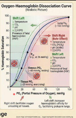
21. Oxygen-Haemoglobin Dissociation Curve
The oxygen-haemoglobin dissociation curve shows the relationship
between the partial pressure of oxygen and the percentage saturation
of haemoglobin with oxygen.
Shape
The curve is sigmoid because oxygen binding to haemoglobin is
cooperative. Binding of one oxygen molecule increases the affinity
of haemoglobin for subsequent oxygen molecules.
Importance
The plateau at high PO₂ helps maintain substantial haemoglobin
saturation in the lungs even when alveolar PO₂ changes moderately.
The steep portion at lower PO₂ facilitates oxygen unloading in tissues.
Right Shift
A rightward shift indicates decreased haemoglobin affinity for oxygen
and therefore enhanced oxygen unloading.
Increased CO₂.
Increased H⁺.
Decreased pH.
Increased temperature.
Increased 2,3-BPG.
Bohr Effect
The Bohr effect refers to the reduction in haemoglobin affinity for
oxygen caused by increased CO₂ and H⁺ concentration. This promotes
oxygen unloading in metabolically active tissues.
Example:
Actively contracting skeletal muscle produces more CO₂, H⁺ and heat.
These conditions shift the oxygen-haemoglobin curve toward the right,
allowing more oxygen to be released to the tissue.
Figure: Oxygen-haemoglobin dissociation curve and factors affecting
haemoglobin oxygen affinity.
22. Haldane Effect
The Haldane effect describes the influence of oxygenation of
haemoglobin on carbon dioxide transport.
Deoxygenated haemoglobin has a greater capacity to bind carbon dioxide
and hydrogen ions than oxygenated haemoglobin. Therefore, in systemic
tissues, deoxygenation of haemoglobin facilitates uptake and transport
of CO₂.
In the lungs, oxygen binding to haemoglobin reduces its affinity
for CO₂ and H⁺, facilitating CO₂ unloading and exhalation.
Tissue
Lungs
Hb becomes deoxygenated and can carry more CO₂ and H⁺.
Hb becomes oxygenated and releases CO₂ and H⁺.
23. Ventilation-Perfusion Relationship
The ventilation-perfusion ratio describes the relationship between
alveolar ventilation and pulmonary blood flow.
V/Q = Alveolar ventilation ÷ Pulmonary perfusion
For the whole lung at rest, the average V/Q ratio is approximately
0.8.
Low V/Q
A low V/Q ratio means that ventilation is inadequate relative to
perfusion. Blood leaving such regions may remain relatively
underoxygenated.
High V/Q
A high V/Q ratio means that ventilation is relatively high compared
with perfusion. A physiologically extreme example is dead space,
where ventilation occurs with little or no effective perfusion.
Importance
Efficient gas exchange requires appropriate matching of ventilation
and perfusion. V/Q mismatch is an important mechanism contributing
to impaired oxygenation in several pulmonary disorders.
24. Regulation of Respiration
Breathing is regulated by respiratory centres in the brainstem,
together with sensory input from chemoreceptors and pulmonary
receptors.
Medulla Oblongata
The medulla contains major neuronal networks involved in generating
the basic respiratory rhythm and coordinating inspiration and expiration.
Pons
Pontine respiratory centres modify and coordinate the respiratory
pattern and contribute to smooth transitions between inspiration
and expiration.
Chemoreceptors
Central Chemoreceptors
Central chemoreceptors respond mainly to changes related to CO₂ and
hydrogen ion concentration in the brain extracellular fluid.
An increase in arterial CO₂ generally increases respiratory drive.
Peripheral Chemoreceptors
Peripheral chemoreceptors are located mainly in the carotid and
aortic bodies. They respond to decreased arterial PO₂ and also
respond to changes in CO₂ and pH.
Basic Feedback Mechanism
↑ CO₂
→
Chemoreceptor stimulation
→
Respiratory centre activation
→
↑ Ventilation
→
↓ CO₂
25. Complete 10-Mark Long Questions
Most important section for examination preparation.
These answers are arranged so that anatomy and physiology can be
connected rather than studied as isolated facts.
LQ 1. Describe the different parts of the respiratory system with their gross and microscopic anatomy and functions.
10 marks
Introduction
The respiratory system is responsible for ventilation and exchange
of oxygen and carbon dioxide. It consists of conducting and
respiratory portions.
Parts
The major parts include nose, nasal cavity, pharynx, larynx,
trachea, bronchi, bronchioles and lungs containing alveoli.
Nose and Nasal Cavity
The nasal cavity is divided by the septum and contains nasal
conchae that increase the surface area and produce turbulence.
Most respiratory regions are lined by pseudostratified ciliated
columnar epithelium containing goblet cells. It filters, warms
and humidifies inspired air.
Pharynx
The pharynx consists of nasopharynx, oropharynx and laryngopharynx.
It conducts air toward the larynx and also participates in swallowing.
Larynx
The larynx is a cartilaginous organ containing vocal folds.
It maintains airway patency, protects the lower airway and
participates in phonation.
Trachea
The trachea is supported by C-shaped hyaline cartilage rings.
Its mucosa contains pseudostratified ciliated columnar epithelium
with goblet cells. Mucociliary activity removes inhaled particles.
Bronchi and Bronchioles
Bronchi contain cartilage plates, smooth muscle and respiratory
epithelium. Bronchioles lack cartilage and glands and contain
relatively prominent smooth muscle.
Lungs and Alveoli
The lungs are paired organs containing a branching bronchial tree
and millions of alveoli. Alveoli are lined mainly by type I
pneumocytes, while type II pneumocytes produce surfactant.
Alveolar macrophages remove particles.
Functions
Ventilation.
Gas exchange.
Oxygen uptake.
Carbon dioxide removal.
Acid-base regulation.
Voice production.
Air filtration and humidification.
Protection against inhaled particles and microorganisms.
Conclusion
The respiratory tract is structurally adapted to conduct air,
protect the lower airway and provide a large, thin surface for
efficient gas exchange.
LQ 2. Describe the gross and microscopic anatomy of lungs and alveoli and explain their functions.
10 marks
Gross Anatomy
The lungs are paired organs located in the thoracic cavity.
The right lung has three lobes, while the left lung has two lobes
and a cardiac notch. Each lung has costal, mediastinal and
diaphragmatic surfaces.
Hilum
The hilum contains structures forming the root of the lung,
including bronchi, pulmonary vessels, lymphatics and nerves.
Microscopic Anatomy
The bronchial tree divides progressively into bronchi, bronchioles,
respiratory bronchioles, alveolar ducts and alveolar sacs.
Alveoli
Alveoli are lined mainly by type I pneumocytes. Type II pneumocytes
produce surfactant. Alveolar macrophages remove foreign particles.
Respiratory Membrane
The respiratory membrane provides a thin barrier between alveolar
air and pulmonary capillary blood. Its small thickness and large
surface area facilitate diffusion.
Functions
Oxygen uptake.
Carbon dioxide elimination.
Maintenance of acid-base balance.
Contribution to pulmonary endocrine and metabolic functions.
Filtration and defense of inhaled materials.
LQ 3. Explain the mechanism of breathing in detail.
10 marks
Introduction
Breathing is the mechanical process by which air moves into and
out of the lungs. It depends on changes in thoracic volume and
pressure.
Forced expiration requires active contraction of internal
intercostal and abdominal muscles.
Role of Surfactant
Pulmonary surfactant reduces alveolar surface tension and helps
maintain alveolar stability, thereby reducing the tendency of
small alveoli to collapse.
Conclusion
Thus, breathing depends on coordinated activity of respiratory
muscles, changes in thoracic volume, pressure gradients and
elastic properties of the lungs.
LQ 4. Explain pulmonary ventilation, alveolar ventilation, lung volumes and capacities.
10 marks
Pulmonary Ventilation
Pulmonary ventilation is the movement of air between atmosphere
and alveoli. It consists of inspiration and expiration.
Minute Ventilation
VE = VT × f
Where VE is minute ventilation, VT is tidal volume and f is
respiratory frequency.
Alveolar Ventilation
VA = (VT − VD) × f
Alveolar ventilation represents the amount of fresh air reaching
gas-exchanging regions per minute.
Lung Volumes
Tidal volume
Inspiratory reserve volume
Expiratory reserve volume
Residual volume
Lung Capacities
Inspiratory capacity
Functional residual capacity
Vital capacity
Total lung capacity
Importance
These measurements provide information about lung function and
help distinguish different patterns of respiratory impairment.
LQ 5. Explain gas exchange between alveoli and blood with special reference to partial pressure of O₂ and CO₂.
10 marks
Introduction
Gas exchange occurs by diffusion across the respiratory membrane
according to partial-pressure gradients.
Oxygen Diffusion
Alveolar PO₂ is approximately 100 mmHg, whereas incoming
pulmonary arterial blood has a PO₂ of approximately 40 mmHg.
Therefore oxygen diffuses from alveoli into pulmonary capillary blood.
Carbon Dioxide Diffusion
Mixed venous blood entering pulmonary capillaries has a PCO₂
of approximately 45 mmHg, while alveolar PCO₂ is approximately
40 mmHg. Therefore CO₂ diffuses from blood into alveoli.
Why Diffusion Occurs
Gases move from regions of higher partial pressure toward regions
of lower partial pressure.
Factors Affecting Diffusion
Partial-pressure gradient.
Surface area.
Membrane thickness.
Gas solubility.
Ventilation-perfusion matching.
Clinical Significance
Thickening of the respiratory membrane or reduction in functional
surface area can impair gas exchange and reduce oxygen transfer.
LQ 6. Explain the transport of oxygen in blood and discuss the oxygen-haemoglobin dissociation curve.
10 marks
Transport of Oxygen
Oxygen is transported in blood in two forms: dissolved in plasma
and bound reversibly to haemoglobin.
Dissolved Oxygen
A small amount is physically dissolved in plasma and determines
the oxygen partial pressure.
Haemoglobin-Bound Oxygen
Most oxygen is transported as oxyhaemoglobin. Each haemoglobin
molecule can bind four oxygen molecules.
Hb + O₂ ⇌ HbO₂
Oxygen Loading
High PO₂ in pulmonary capillaries promotes oxygen loading onto
haemoglobin.
Oxygen Unloading
Lower PO₂ in tissues promotes oxygen release.
Increased CO₂, H⁺, temperature and 2,3-BPG further promote unloading.
Dissociation Curve
The oxygen-haemoglobin dissociation curve is sigmoid because
haemoglobin shows cooperative oxygen binding.
Right Shift
Increased CO₂, increased H⁺, increased temperature and increased
2,3-BPG shift the curve to the right and promote oxygen unloading.
Bohr Effect
The Bohr effect describes the reduction in haemoglobin affinity
for oxygen caused by increased CO₂ and H⁺ concentration,
facilitating oxygen delivery to metabolically active tissues.
LQ 7. Describe the transport of carbon dioxide in blood in detail.
10 marks
Introduction
Carbon dioxide produced by cellular metabolism is transported
from tissues to lungs in three major forms.
1. Dissolved CO₂
A small proportion is physically dissolved in plasma.
2. Carbamino Compounds
CO₂ binds to amino groups of proteins, particularly haemoglobin,
forming carbaminohaemoglobin.
3. Bicarbonate
Most CO₂ is transported as bicarbonate ions.
In red blood cells, carbonic anhydrase catalyses:
CO₂ + H₂O ⇌ H₂CO₃ ⇌ H⁺ + HCO₃⁻
Bicarbonate leaves the red cell in exchange for chloride ions,
producing the chloride shift.
At the Lungs
Oxygenation of haemoglobin promotes release of hydrogen ions
and CO₂. Bicarbonate re-enters red blood cells and is converted
back to CO₂, which diffuses into alveoli and is exhaled.
Importance
CO₂ transport is closely associated with maintenance of
acid-base balance because the bicarbonate buffer system is
an important extracellular buffering system.
LQ 8. Explain the mechanisms of external and internal respiration.
10 marks
External Respiration
External respiration occurs between alveolar air and pulmonary
capillary blood.
Oxygen diffuses from alveoli into blood because alveolar PO₂
is higher than venous blood PO₂.
Carbon dioxide diffuses from pulmonary capillary blood into
alveoli because venous PCO₂ is higher than alveolar PCO₂.
Internal Respiration
Internal respiration occurs between systemic capillary blood
and tissue cells.
Oxygen moves from systemic blood to tissues because tissue PO₂
is lower than arterial blood PO₂.
Carbon dioxide produced by tissues diffuses into systemic blood.
Overall Sequence
Atmosphere
→
Alveoli
→
Pulmonary Blood
→
Systemic Blood
→
Tissues
LQ 9. Explain the regulation of respiration.
10 marks
Introduction
Respiration is regulated to maintain adequate oxygen delivery,
carbon dioxide elimination and acid-base balance.
Medullary Centres
Neuronal networks in the medulla generate and coordinate the
basic respiratory rhythm.
Pontine Centres
Pontine centres modify the respiratory pattern and coordinate
transitions between inspiration and expiration.
Central Chemoreceptors
Central chemoreceptors respond primarily to changes associated
with CO₂ and hydrogen ion concentration in the brain extracellular
fluid.
Peripheral Chemoreceptors
Carotid and aortic bodies respond to decreased arterial PO₂ and
also to changes in CO₂ and pH.
Response to Increased CO₂
Increased arterial CO₂ increases respiratory drive, increasing
ventilation. Increased ventilation removes CO₂ and tends to
restore arterial CO₂ toward normal.
Conclusion
Respiratory regulation depends on coordinated activity of brainstem
centres, chemoreceptors and respiratory muscles.
LQ 10. Explain ventilation-perfusion relationship and its importance in gas exchange.
10 marks
Definition
Ventilation refers to movement of air into alveoli, whereas
perfusion refers to blood flow through pulmonary capillaries.
V/Q = Alveolar ventilation / Pulmonary perfusion
The average whole-lung V/Q ratio is approximately 0.8 at rest.
Low V/Q
When perfusion is relatively high compared with ventilation,
blood may leave the affected region with reduced oxygenation.
High V/Q
When ventilation exceeds perfusion, some inspired air contributes
relatively little to effective gas exchange.
Dead Space
Ventilation of regions with little or no effective perfusion
represents physiological dead-space ventilation.
Importance
Efficient oxygen uptake and carbon dioxide removal require
appropriate matching of alveolar ventilation with pulmonary
perfusion.
LQ 11. Describe the microscopic anatomy of the respiratory tract from trachea to alveoli.
10 marks
Trachea
The trachea is lined by pseudostratified ciliated columnar
epithelium with goblet cells. Its wall contains submucosal
glands and C-shaped hyaline cartilage.
Bronchi
Bronchi retain respiratory epithelium, smooth muscle, glands and
cartilage plates.
Bronchioles
Bronchioles lack cartilage and submucosal glands. Their epithelium
becomes simple columnar or cuboidal and smooth muscle is relatively
prominent.
Terminal Bronchioles
Terminal bronchioles are lined predominantly by simple cuboidal
epithelium and contain club cells.
Respiratory Bronchioles
Respiratory bronchioles contain alveoli in their walls and represent
the beginning of the respiratory zone.
Alveoli
Alveoli are lined mainly by type I pneumocytes. Type II pneumocytes
produce surfactant and alveolar macrophages provide defense.
Functional Correlation
The progressive reduction in epithelial thickness and structural
barriers toward the alveoli reflects the increasing importance
of efficient gas exchange.
LQ 12. Explain the integrated process of respiration from atmospheric air to tissue cells.
10 marks
Step 1: Pulmonary Ventilation
Atmospheric air enters the lungs during inspiration and leaves
during expiration.
Step 2: Alveolar Gas Exchange
Oxygen diffuses from alveoli to pulmonary blood and carbon dioxide
diffuses in the opposite direction.
Step 3: Oxygen Transport
Most oxygen binds reversibly to haemoglobin and is transported
through systemic circulation.
Step 4: Tissue Exchange
Oxygen diffuses from systemic capillary blood to tissues.
Step 5: Carbon Dioxide Uptake
Carbon dioxide generated by cellular metabolism diffuses into
blood and is transported mainly as bicarbonate.
Step 6: Pulmonary Return
Venous blood reaches pulmonary capillaries, where carbon dioxide
is unloaded and oxygen is taken up.
Atmosphere
→
Ventilation
→
Alveoli
→
Blood
→
Tissues
→
CO₂ Return
→
Lungs
26. Separate 5-Mark Preparation
For 5 marks:
Write a short introduction followed by 5–7 well-organized points.
Do not write only definitions when the question asks for anatomy
or mechanism.
1. Functions of Respiratory System
Oxygen uptake.
Carbon dioxide removal.
Maintenance of acid-base balance.
Voice production.
Air filtration, warming and humidification.
Defense against inhaled particles.
2. Conducting and Respiratory Zones
The conducting zone extends from nose to terminal bronchioles and
conducts, filters, warms and humidifies air. The respiratory zone
consists of respiratory bronchioles, alveolar ducts and alveoli and
is responsible for gas exchange.
3. Histology of Trachea
Pseudostratified ciliated columnar epithelium.
Goblet cells.
Submucosal seromucous glands.
C-shaped hyaline cartilage.
Trachealis muscle posteriorly.
4. Histology of Alveoli
Type I pneumocytes for gas exchange.
Type II pneumocytes produce surfactant.
Alveolar macrophages provide defense.
Thin respiratory membrane.
Dense pulmonary capillary network.
5. Mechanism of Inspiration
Diaphragm contracts.
External intercostals contract.
Thoracic volume increases.
Alveolar pressure decreases.
Air enters the lungs.
6. Mechanism of Expiration
Inspiratory muscles relax.
Thoracic volume decreases.
Elastic recoil of lungs occurs.
Alveolar pressure increases.
Air leaves lungs.
7. Gas Exchange
Oxygen diffuses from alveoli to blood because alveolar PO₂ is higher
than venous blood PO₂. Carbon dioxide diffuses from blood to alveoli
because venous PCO₂ is higher than alveolar PCO₂.
8. Partial Pressure
Partial pressure is the pressure exerted by an individual gas in a
mixture. At sea level, atmospheric PO₂ is approximately 159 mmHg,
while alveolar PO₂ is approximately 100 mmHg and alveolar PCO₂ is
approximately 40 mmHg.
9. Oxygen Transport
Oxygen is transported mainly bound to haemoglobin and a small amount
dissolved in plasma. Haemoglobin binds oxygen reversibly to form
oxyhaemoglobin.
10. Carbon Dioxide Transport
Dissolved in plasma.
As carbamino compounds.
Mostly as bicarbonate.
11. Bohr Effect
Increased CO₂ and H⁺ decrease haemoglobin affinity for oxygen and
promote oxygen unloading in tissues.
12. Haldane Effect
Deoxygenated haemoglobin carries more CO₂ and H⁺, while oxygenation
of haemoglobin in lungs promotes CO₂ unloading.
13. Surfactant
Pulmonary surfactant is produced by type II pneumocytes. It lowers
alveolar surface tension, increases lung compliance and reduces
the tendency of alveoli to collapse.
14. V/Q Ratio
V/Q represents the relationship between alveolar ventilation and
pulmonary perfusion. The average whole-lung value is approximately
0.8 at rest.
15. Regulation of Respiration
Respiration is regulated by brainstem respiratory centres and
chemoreceptors. Increased CO₂ generally stimulates ventilation,
while peripheral chemoreceptors also respond strongly to low arterial
PO₂.
27. Separate 2-Mark Preparation
Question
Answer
Define respiration.
Respiration is the integrated process involving ventilation,
gas exchange, transport of respiratory gases and tissue exchange.
Name the major parts of respiratory system.
Nose, nasal cavity, pharynx, larynx, trachea, bronchi,
bronchioles and lungs.
What is pulmonary ventilation?
Movement of air between atmosphere and alveoli.
What is external respiration?
Exchange of O₂ and CO₂ between alveolar air and pulmonary blood.
What is internal respiration?
Exchange of gases between systemic blood and tissue cells.
What is tidal volume?
Volume of air inspired or expired during normal quiet breathing;
approximately 500 mL in a typical adult.
What is residual volume?
Volume of air remaining in the lungs after maximal expiration.
What is alveolar ventilation?
Volume of fresh air reaching functioning respiratory regions per minute.
What is dead space?
Portion of ventilation that does not participate directly in gas exchange.
Name the types of pneumocytes.
Type I and Type II pneumocytes.
Function of Type II pneumocytes?
They synthesize and secrete pulmonary surfactant.
What is pulmonary surfactant?
A surface-active material that reduces alveolar surface tension
and helps prevent alveolar collapse.
What is respiratory membrane?
The thin alveolar-capillary barrier across which O₂ and CO₂ diffuse.
What is partial pressure?
The pressure contributed by an individual gas in a mixture.
Normal alveolar PO₂?
Approximately 100 mmHg at sea level.
Normal alveolar PCO₂?
Approximately 40 mmHg.
Normal mixed venous PO₂?
Approximately 40 mmHg.
Normal mixed venous PCO₂?
Approximately 45 mmHg.
How is most oxygen transported?
Bound reversibly to haemoglobin as oxyhaemoglobin.
How is most CO₂ transported?
Mainly as bicarbonate ions.
What is Bohr effect?
Decreased haemoglobin affinity for O₂ due to increased CO₂ and H⁺,
promoting oxygen unloading.
What is Haldane effect?
Deoxygenated haemoglobin has greater capacity to carry CO₂ and H⁺,
while oxygenation promotes CO₂ release.
What is chloride shift?
Exchange of bicarbonate leaving red cells for chloride ions entering
them during CO₂ transport.
What is V/Q ratio?
Ratio of alveolar ventilation to pulmonary perfusion; average
whole-lung value is approximately 0.8.
What is respiratory zone?
Region consisting of respiratory bronchioles, alveolar ducts,
alveolar sacs and alveoli where gas exchange occurs.
What is conducting zone?
Air-conducting portion extending from nose to terminal bronchioles.
Name the respiratory muscles.
Diaphragm and intercostal muscles are the major muscles;
accessory muscles assist during increased respiratory effort.
What is the main muscle of inspiration?
Diaphragm.
What is the function of alveolar macrophages?
Removal of inhaled particles, microorganisms and cellular debris.
28. Quick Revision
Respiratory Tract Sequence
Nose
→
Pharynx
→
Larynx
→
Trachea
→
Bronchi
→
Bronchioles
→
Alveoli
Gas Exchange
Gas
Direction in lungs
Main reason
O₂
Alveoli → Blood
Higher alveolar PO₂
CO₂
Blood → Alveoli
Higher blood PCO₂
Important Partial Pressures
Location
PO₂
PCO₂
Atmosphere
~159 mmHg
~0.3 mmHg
Alveoli
~100 mmHg
~40 mmHg
Mixed venous blood
~40 mmHg
~45 mmHg
Oxygen Transport
Main Form
Oxyhaemoglobin
Curve
Sigmoid due to cooperative binding
Right Shift
↑ CO₂, ↑ H⁺, ↑ temperature, ↑ 2,3-BPG
Carbon Dioxide Transport
1
Dissolved CO₂
2
Carbamino compounds
3
Bicarbonate
Breathing Mechanism
Inspiration
Expiration
Diaphragm contracts
Diaphragm relaxes
Thoracic volume increases
Thoracic volume decreases
Alveolar pressure decreases
Alveolar pressure increases
Air enters lungs
Air leaves lungs
One-line memory sequence:
Ventilation → Alveolar gas exchange → Blood transport →
Tissue gas exchange → Cellular respiration
Important Exam Points
For an anatomy LQ, do not stop at naming respiratory organs.
Explain their gross anatomy + microscopic anatomy + functions.
Alveoli are the principal site of gas exchange.
Type I pneumocytes form most of the gas-exchange surface.
Type II pneumocytes produce surfactant.
Inspiration is normally an active muscular process.
Quiet expiration is mainly passive because of elastic recoil.
Air moves because of pressure gradients created by changes in
thoracic volume.
Oxygen moves from alveoli to blood because alveolar PO₂ exceeds
pulmonary venous blood PO₂ during equilibration.
Carbon dioxide moves from pulmonary blood to alveoli because
pulmonary capillary PCO₂ exceeds alveolar PCO₂.
Most oxygen is transported bound to haemoglobin.
Most carbon dioxide is transported as bicarbonate.
Carbonic anhydrase greatly accelerates bicarbonate formation
inside red blood cells.
The Bohr effect facilitates oxygen unloading in metabolically
active tissues.
The Haldane effect facilitates carbon dioxide uptake in tissues
and unloading in lungs.
V/Q matching is essential for efficient pulmonary gas exchange.
Increased arterial CO₂ generally increases ventilation through
respiratory control mechanisms.
Diagrams You Should Practice for 10-Mark Questions
1. Complete Respiratory System
Label nasal cavity, pharynx, larynx, trachea, bronchi,
bronchioles and lungs.
2. Lung Anatomy
Show right and left lungs, lobes, fissures, hilum and major bronchi.
3. Alveolus
Label alveolar space, Type I pneumocyte, Type II pneumocyte,
macrophage, capillary and respiratory membrane.
4. Mechanism of Breathing
Draw inspiration and expiration showing diaphragm movement,
thoracic volume and pressure changes.
5. Gas Exchange
Show O₂ movement from alveolus to blood and CO₂ movement from
blood to alveolus with partial pressures.
6. O₂-Hb Curve
Draw sigmoid curve and indicate right shift, Bohr effect and
major factors affecting oxygen affinity.
7. CO₂ Transport
Show dissolved CO₂, carbaminohaemoglobin and bicarbonate formation,
including chloride shift.
8. V/Q Relationship
Explain normal ventilation-perfusion matching and the concepts
of low V/Q and high V/Q.
Selected Clinical Correlations
Asthma
Asthma involves variable airway narrowing and increased airway
resistance. Bronchial smooth-muscle constriction, inflammation
and mucus production can impair airflow.
Pneumonia
Infection and inflammatory material within alveoli can interfere
with effective ventilation and gas exchange.
Pulmonary Edema
Accumulation of fluid within the pulmonary interstitium or alveolar
spaces can increase diffusion distance and impair oxygen transfer.
Emphysema
Destruction of alveolar walls reduces the effective surface area
available for gas exchange and alters elastic recoil.
Pulmonary Embolism
Obstruction of pulmonary blood flow creates regions that may be
ventilated but poorly perfused, producing a high V/Q state.
How to Write a 10-Mark Respiratory LQ
Recommended Pattern
Introduction / definition
Classification or parts
Gross anatomy
Microscopic anatomy
Functions
Physiological mechanism where applicable
Relevant diagram
Important physiological values where applicable
Clinical significance where relevant
Conclusion
For physiology-heavy questions such as gas exchange, oxygen transport
and breathing mechanism, emphasize the sequence of events and use
pressure values and equations where appropriate. For anatomy-heavy
questions, emphasize labelled structure, histology and functional
correlation.