Chapter Navigation
Overview Parts & Functions Neuron Neuroglia Microscopic Anatomy Brain Spinal Cord Action Potential Nerve Impulse Synapse Ventricular System CSF Meninges Reflex Arc Autonomic Nervous System 10 Mark LQs 5 Mark 2 MarkMB 551 Syllabus Coverage
The Nervous System is one of the most important chapters of Human Anatomy and Physiology because it combines structural anatomy with functional physiology. In examination, questions may be asked from the gross anatomy of the brain and spinal cord, microscopic structure of nervous tissue, neurons and neuroglia, action potential, conduction of nerve impulses, synaptic transmission, ventricular system, cerebrospinal fluid and meninges. Therefore, this chapter is prepared as a complete long-question chapter.
- Parts and functions of the nervous system
- Gross structure and microscopic anatomy of the nervous system
- Ventricular system and cerebrospinal fluid
- Meninges
- Neuron structure and classification
- Neuroglia
- Resting membrane potential
- Action potential
- Depolarization and repolarization
- Refractory periods
- Conduction of nerve impulse
- Saltatory and continuous conduction
- Synaptic transmission
- Reflex arc
- Autonomic nervous system
- Blood-brain barrier
- Functional organization of CNS and PNS
1. Nervous System: General Overview
The nervous system is the highly specialized communication and control system of the body that receives sensory information, processes and integrates the information, and produces appropriate motor or autonomic responses. It coordinates the activities of different organs and helps maintain homeostasis.
The nervous system allows the body to detect changes in the internal and external environment, transmit information rapidly, interpret sensory signals, generate appropriate responses and coordinate complex functions such as movement, sensation, learning, memory, emotion and regulation of visceral organs.
Major Functions
| Function | Explanation |
|---|---|
| Sensory function | Receptors detect changes such as touch, pain, temperature, pressure, light, sound, chemical substances and changes in internal body conditions. |
| Integration | Sensory information is analyzed and interpreted mainly within the CNS, allowing the body to determine an appropriate response. |
| Motor function | Motor commands are transmitted to skeletal muscles, smooth muscles, cardiac muscle and glands. |
| Homeostasis | The nervous system works with the endocrine system to regulate temperature, blood pressure, respiration, digestion, fluid balance and other functions. |
| Higher functions | The brain is responsible for consciousness, thinking, memory, learning, language, emotions and voluntary behavior. |
| Protection | Reflex responses provide rapid protective responses to potentially harmful stimuli. |
2. Parts of the Nervous System
Anatomically, the nervous system is divided into the central nervous system and peripheral nervous system. Functionally, the peripheral nervous system is further divided into somatic and autonomic divisions.
Central Nervous System
- Brain
- Spinal cord
- Main center for integration and control
- Protected by skull, vertebral column, meninges and CSF
Peripheral Nervous System
- Cranial nerves
- Spinal nerves
- Peripheral ganglia
- Receptors and peripheral nerve endings
Functional Classification
| Division | Major Components | Main Function |
|---|---|---|
| Somatic nervous system | Sensory and motor pathways associated mainly with skin, joints and skeletal muscles | Conscious sensation and voluntary control of skeletal muscles |
| Autonomic nervous system | Sympathetic, parasympathetic and enteric divisions | Regulation of smooth muscle, cardiac muscle and glands |
| Sympathetic | Thoracolumbar outflow | Supports responses associated with increased activity and stress |
| Parasympathetic | Craniosacral outflow | Supports rest, digestion and energy conservation |
| Enteric nervous system | Myenteric and submucosal plexuses | Coordinates gastrointestinal motility and secretion |

3. Neuron: Structural and Functional Unit of Nervous System
The neuron is the structural and functional unit of the nervous system. It is a specialized excitable cell capable of receiving stimuli, generating electrical signals and transmitting information to another neuron, muscle or gland.
Basic Structure of a Neuron
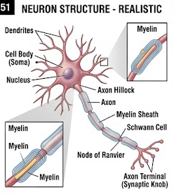
1. Cell Body or Soma
The cell body contains the nucleus and most of the metabolic machinery required for maintenance of the neuron. It contains abundant rough endoplasmic reticulum and ribosomes, collectively visible as Nissl substance. The soma also contains mitochondria, Golgi apparatus, lysosomes, cytoskeletal elements and other organelles.
The nucleus is usually large and euchromatic, reflecting high metabolic activity. The nucleolus is prominent. The cell body maintains the metabolic and synthetic functions of the neuron.
2. Dendrites
Dendrites are usually short, branched cytoplasmic processes that receive signals from sensory receptors or other neurons. Their branching increases the surface area available for synaptic contacts.
3. Axon
The axon is a specialized process that conducts electrical signals away from the neuronal cell body. A neuron generally has one axon, although the axon may branch extensively.
The axon originates from the axon hillock and continues into the initial segment. The axon hillock and initial segment are important regions for the initiation of action potentials because they contain a high density of voltage-gated ion channels.
4. Myelin Sheath
Myelin is a lipid-rich insulating covering around many axons. In the peripheral nervous system it is produced by Schwann cells, while in the central nervous system it is produced by oligodendrocytes.
Myelination increases the speed and efficiency of nerve impulse conduction. The myelin sheath is interrupted at regular intervals by nodes of Ranvier.
5. Nodes of Ranvier
Nodes of Ranvier are small gaps between adjacent myelin segments. Voltage-gated sodium and potassium channels are concentrated at these nodes. In myelinated axons, the action potential is regenerated at the nodes and appears to jump from node to node. This is called saltatory conduction.
6. Axon Terminals
The terminal branches of an axon end in synaptic terminals or boutons. They contain synaptic vesicles filled with neurotransmitters. Arrival of an action potential at the terminal causes voltage-gated calcium channels to open and triggers neurotransmitter release.
Classification of Neurons
| Type | Structure | Example / Function |
|---|---|---|
| Unipolar / pseudounipolar | Single process that divides into peripheral and central branches | Common sensory neurons of spinal dorsal root ganglia |
| Bipolar | One dendrite and one axon | Retina, olfactory and vestibulocochlear pathways |
| Multipolar | One axon and many dendrites | Most motor neurons and many CNS neurons |
4. Neuroglia
Neuroglia are non-neuronal cells that support, protect, nourish and maintain the environment of neurons. Unlike mature neurons, many glial cells retain the ability to divide.
| Glial cell | Location | Major functions |
|---|---|---|
| Astrocytes | CNS | Structural support, ion and neurotransmitter regulation, metabolic support, participation in blood-brain barrier, repair and glial scar formation. |
| Oligodendrocytes | CNS | Form myelin around CNS axons. |
| Microglia | CNS | Resident immune/phagocytic cells of CNS; remove damaged cells and debris. |
| Ependymal cells | Ventricles and central canal | Line ventricular spaces and participate in CSF production and circulation. |
| Schwann cells | PNS | Form myelin around peripheral axons and support regeneration. |
| Satellite cells | Peripheral ganglia | Surround neuronal cell bodies and regulate their local environment. |
5. Microscopic Anatomy of Nervous Tissue
Gray Matter
Gray matter contains neuronal cell bodies, dendrites, axons, neuroglia and synaptic connections. In the brain, gray matter forms the cerebral and cerebellar cortex and is also present in deeper nuclei. In the spinal cord, gray matter occupies the central region.
White Matter
White matter contains predominantly myelinated axons arranged in bundles or tracts. The lipid-rich myelin gives white matter its lighter appearance. In the brain, white matter is mainly located deep to the cortex, whereas in the spinal cord it surrounds the central gray matter.
Microscopic Structure of Peripheral Nerves
Peripheral nerves contain axons surrounded by connective tissue coverings. Individual nerve fibers are surrounded by endoneurium. Groups of nerve fibers form fascicles surrounded by perineurium. The entire nerve is surrounded by epineurium.
| Layer | Surrounds | Importance |
|---|---|---|
| Endoneurium | Individual nerve fiber | Fine connective tissue support around individual axons. |
| Perineurium | Nerve fascicle | Provides mechanical protection and contributes to the blood-nerve barrier. |
| Epineurium | Entire nerve | Strong outer connective tissue covering containing blood vessels. |

6. Brain: Gross and Microscopic Anatomy
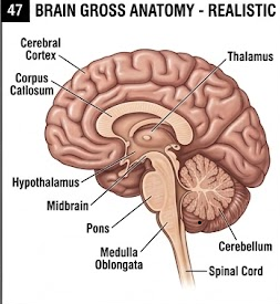
The brain is the major control center of the central nervous system. It is located within the cranial cavity and is protected by the skull, meninges and cerebrospinal fluid.
Major Divisions of Brain
| Division | Major structures | Important functions |
|---|---|---|
| Forebrain | Cerebrum, diencephalon | Consciousness, sensation, voluntary movement, cognition, memory, emotion and endocrine/autonomic regulation. |
| Midbrain | Mesencephalon | Visual and auditory reflexes, motor pathways and integration of sensory information. |
| Hindbrain | Pons, medulla and cerebellum | Coordination, posture, balance, cardiovascular and respiratory control and relay functions. |
Cerebrum
The cerebrum is the largest portion of the brain and consists of right and left cerebral hemispheres. The hemispheres are connected mainly by the corpus callosum. The cerebral surface is folded into gyri separated by sulci, greatly increasing the cortical surface area.
The cerebral cortex consists primarily of gray matter and is organized into functional areas associated with motor activity, somatic sensation, vision, hearing, language and higher mental functions.
Major Lobes of Cerebrum
| Lobe | Major functions |
|---|---|
| Frontal lobe | Voluntary motor control, planning, personality, judgment, speech production and executive functions. |
| Parietal lobe | Somatic sensory processing, spatial orientation and integration of sensory information. |
| Temporal lobe | Hearing, memory, language comprehension and aspects of emotion. |
| Occipital lobe | Visual processing. |
| Insula | Visceral sensation, taste and integration of internal body signals. |
Cerebellum
The cerebellum is located posterior to the brainstem. It consists of two hemispheres connected by the vermis. Its major functions include coordination of voluntary movements, maintenance of posture, balance and motor learning.
Brainstem
The brainstem consists of the midbrain, pons and medulla oblongata. It contains important ascending and descending pathways and nuclei involved in cranial nerve functions. The medulla contains important autonomic centers involved in cardiovascular and respiratory regulation.
Diencephalon
The diencephalon includes the thalamus, hypothalamus and associated structures. The thalamus acts as an important relay and processing center for many sensory pathways. The hypothalamus regulates homeostasis, temperature, appetite, thirst, endocrine activity and autonomic functions.
Microscopic Structure of Cerebral Cortex
The cerebral cortex is composed of gray matter containing neuronal cell bodies, dendrites, axons, glial cells and blood vessels. The neocortex is classically organized into six cellular layers. The relative proportions and neuronal populations vary between cortical regions.
| Layer | General characteristic |
|---|---|
| I – Molecular layer | Contains relatively few neuronal cell bodies and many dendritic/axonal processes. |
| II – External granular layer | Contains small neurons and participates in intracortical connections. |
| III – External pyramidal layer | Contains pyramidal neurons involved in corticocortical connections. |
| IV – Internal granular layer | Important recipient of many thalamocortical sensory inputs. |
| V – Internal pyramidal layer | Contains large pyramidal neurons contributing to descending motor pathways. |
| VI – Multiform layer | Contains diverse neuronal types and contributes to corticothalamic connections. |
Microscopic Structure of Cerebellar Cortex
The cerebellar cortex has three layers: molecular layer, Purkinje cell layer and granular layer. Purkinje cells are large neurons whose axons provide the major output of the cerebellar cortex.
7. Spinal Cord: Gross and Microscopic Anatomy
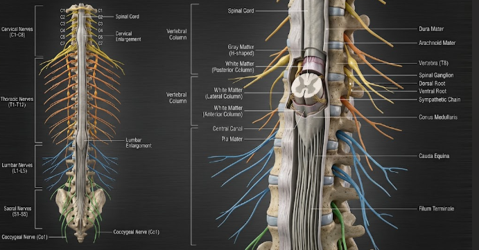
The spinal cord is a cylindrical structure extending from the medulla oblongata through the vertebral canal. It provides a major pathway between the brain and peripheral tissues and also acts as an important center for reflex activity.
Gross Features
- Extends from the medulla to approximately the level of L1–L2 vertebrae in the adult.
- Has cervical and lumbosacral enlargements.
- Ends as the conus medullaris.
- Filum terminale extends inferiorly from the conus.
- Cauda equina consists of spinal nerve roots descending within the vertebral canal.
- Spinal nerves arise from the cord through dorsal and ventral roots.
Dorsal and Ventral Roots
The dorsal root carries sensory information toward the spinal cord and contains the dorsal root ganglion. The ventral root carries motor output away from the spinal cord.
Microscopic Structure
In cross-section, the spinal cord has central gray matter surrounded by white matter. The gray matter has an approximate butterfly or H-shaped appearance.
| Region | Major function |
|---|---|
| Dorsal horn | Receives and processes sensory information. |
| Ventral horn | Contains motor neurons supplying skeletal muscles. |
| Lateral horn | Contains autonomic neurons, particularly prominent in thoracic and upper lumbar levels. |
| White matter | Contains ascending sensory and descending motor pathways. |
| Central canal | Small central channel lined by ependymal cells and associated with the CSF system. |
8. Action Potential: Detailed Mechanism
An action potential is a rapid, transient and propagated change in the membrane potential of an excitable cell produced by the sequential opening and closing of voltage-gated ion channels.
Resting Membrane Potential
When a neuron is not conducting an action potential, its plasma membrane has a resting membrane potential. In a typical neuron this is approximately −70 mV, although the exact value varies between cells.
The resting membrane potential results mainly from unequal distribution of ions across the membrane, selective membrane permeability, potassium leak channels and the sodium-potassium pump.
Sodium-Potassium Pump
The Na+/K+-ATPase uses ATP to transport three Na+ ions out of the cell and two K+ ions into the cell during each cycle. It maintains the ion gradients necessary for normal neuronal excitability.
Threshold
A sufficiently strong depolarizing stimulus can bring the membrane to a threshold level. Once threshold is reached, voltage-gated sodium channels open rapidly and initiate an action potential.
Phases of Action Potential
| Phase | Ion channel events | Membrane change |
|---|---|---|
| Resting state | Voltage-gated Na+ and K+ channels mostly closed; leak channels active | Approximately −70 mV |
| Threshold | Sufficient depolarization activates voltage-gated Na+ channels | Action potential initiated |
| Depolarization | Rapid Na+ influx | Membrane potential becomes less negative and may become positive |
| Repolarization | Na+ channels inactivate; K+ channels open | Membrane potential becomes negative again |
| Hyperpolarization | K+ channels remain open temporarily | Membrane becomes more negative than resting level |
| Return to resting state | K+ channels close; ionic gradients are maintained by leak pathways and Na+/K+-ATPase | Resting membrane potential restored |
All-or-None Principle
Once threshold is reached, an action potential occurs with a relatively stereotyped amplitude. Increasing the strength of a suprathreshold stimulus does not normally increase the amplitude of an individual action potential. Instead, stimulus intensity can be represented by changes in firing frequency and recruitment of neurons.
Refractory Period
During the absolute refractory period, another action potential cannot be generated regardless of stimulus strength because voltage-gated sodium channels are either open or inactivated.
During the relative refractory period, another action potential can occur but requires a stronger-than-normal stimulus because some sodium channels have recovered while potassium conductance remains elevated.
9. Nerve Impulse and Its Conduction
A nerve impulse is the propagated electrical signal generated by an action potential and transmitted along an axon. The mechanism differs according to whether the axon is myelinated or unmyelinated.
Continuous Conduction
In an unmyelinated axon, every adjacent segment of the membrane undergoes depolarization and repolarization. The action potential therefore appears to travel continuously along the entire axonal membrane.
Saltatory Conduction
In a myelinated axon, myelin acts as an electrical insulator. Voltage-gated ion channels are concentrated at the nodes of Ranvier. Local current spreads rapidly beneath the myelin from one node to the next, allowing the action potential to be regenerated at successive nodes.
Factors Affecting Conduction Velocity
- Myelination: increases conduction velocity.
- Axon diameter: larger axons generally conduct faster.
- Temperature: within physiological limits, temperature affects channel kinetics and conduction.
- Membrane properties: resistance and capacitance influence the speed of electrical propagation.
10. Synapse and Synaptic Transmission
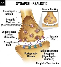
A synapse is a specialized junction through which a neuron communicates with another neuron or an effector cell such as a muscle or gland.
Components of Chemical Synapse
- Presynaptic terminal
- Synaptic vesicles containing neurotransmitter
- Presynaptic membrane
- Synaptic cleft
- Postsynaptic membrane
- Postsynaptic receptors
Mechanism of Chemical Synaptic Transmission
When an action potential reaches the presynaptic terminal, voltage-gated calcium channels open. Calcium enters the terminal and promotes fusion of synaptic vesicles with the presynaptic membrane. Neurotransmitter is released into the synaptic cleft by exocytosis. The neurotransmitter then binds to receptors on the postsynaptic membrane and alters ion permeability or activates intracellular signaling pathways.
Examples of Neurotransmitters
| Neurotransmitter | Important roles |
|---|---|
| Acetylcholine | Neuromuscular transmission, autonomic signaling and CNS functions. |
| Glutamate | Major excitatory neurotransmitter in CNS. |
| GABA | Major inhibitory neurotransmitter in brain. |
| Glycine | Important inhibitory neurotransmitter, especially in spinal cord. |
| Dopamine | Movement, reward, motivation and endocrine regulation. |
| Serotonin | Mood, sleep, appetite and several CNS functions. |
| Noradrenaline | Attention, arousal and sympathetic neurotransmission. |
11. Ventricular System of the Brain
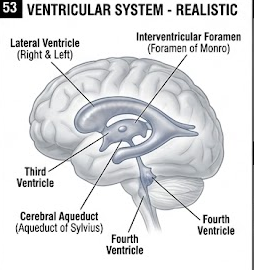
The ventricular system is a series of interconnected cavities within the brain that contain cerebrospinal fluid. The ventricles are lined largely by ependymal cells.
Four Main Ventricles
- Right lateral ventricle
- Left lateral ventricle
- Third ventricle
- Fourth ventricle
CSF Flow Through Ventricular System
Lateral Ventricles
The paired lateral ventricles are located within the cerebral hemispheres. Each communicates with the third ventricle through the interventricular foramen.
Third Ventricle
The third ventricle is a narrow cavity located in the diencephalon between the thalami. It communicates with the fourth ventricle through the cerebral aqueduct.
Cerebral Aqueduct
The cerebral aqueduct is a narrow channel passing through the midbrain. It connects the third and fourth ventricles.
Fourth Ventricle
The fourth ventricle lies between the pons and medulla anteriorly and the cerebellum posteriorly. It communicates with the subarachnoid space through the median and lateral apertures.
12. Cerebrospinal Fluid (CSF)
Cerebrospinal fluid is a clear, relatively low-protein fluid that circulates within the brain ventricles and subarachnoid space around the brain and spinal cord.
Formation of CSF
Most CSF is produced by the choroid plexuses within the ventricles. The choroid plexus contains specialized capillaries and epithelial cells derived from ependymal cells. CSF formation involves selective transport of ions and water from blood into the ventricular system.
Circulation of CSF
CSF flows from the lateral ventricles through the interventricular foramina to the third ventricle. It then passes through the cerebral aqueduct to the fourth ventricle and enters the subarachnoid space through openings of the fourth ventricle.
Absorption of CSF
CSF is mainly returned to the venous circulation through arachnoid granulations into dural venous sinuses. The movement of CSF into the venous system depends on pressure gradients and specialized arachnoid structures.
Functions of CSF
- Provides mechanical protection to the brain and spinal cord.
- Provides buoyancy, reducing the effective weight of the brain.
- Helps maintain a stable extracellular environment for neurons.
- Participates in transport of substances and removal of metabolic waste.
- Helps distribute certain signaling molecules.
13. Meninges
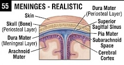
The meninges are three connective tissue coverings surrounding the brain and spinal cord. From outer to inner they are dura mater, arachnoid mater and pia mater.
1. Dura Mater
The dura mater is the tough outer meningeal layer. In the cranial cavity it consists of periosteal and meningeal layers. Dural folds form partitions between major parts of the brain and help limit displacement of brain tissue.
Important dural folds include the falx cerebri, tentorium cerebelli, falx cerebelli and diaphragma sellae.
2. Arachnoid Mater
The arachnoid mater is a thin membrane located beneath the dura. It does not closely follow every contour of the brain. Trabeculae extend from the arachnoid toward the pia and cross the subarachnoid space.
3. Pia Mater
The pia mater is a delicate vascular connective tissue membrane closely attached to the surface of the brain and spinal cord. It follows the contours of the nervous tissue.
Subarachnoid Space
The subarachnoid space lies between arachnoid and pia mater. It contains CSF, blood vessels and connective tissue trabeculae. It therefore provides a fluid-filled protective environment around the CNS.
| Layer | Position | Major feature |
|---|---|---|
| Dura mater | Outer | Strong protective connective tissue layer. |
| Arachnoid mater | Middle | Thin membrane with trabeculae; forms boundary of subarachnoid space. |
| Pia mater | Inner | Closely follows surface of brain and spinal cord. |
14. Blood-Brain Barrier
The blood-brain barrier is a specialized structural and functional barrier that regulates movement of substances from the blood into the extracellular environment of the CNS.
Major Components
- Brain capillary endothelial cells joined by tight junctions.
- Basement membrane.
- Pericytes.
- Astrocytic end-feet surrounding the capillaries.
The blood-brain barrier restricts entry of many large molecules and potentially harmful substances while permitting regulated transport of essential nutrients such as glucose. Lipid-soluble substances and gases such as oxygen and carbon dioxide can cross relatively readily.
15. Reflex Action and Reflex Arc
A reflex is a rapid, automatic response to a stimulus that occurs through a neural pathway called a reflex arc.
Components of Reflex Arc
Types of Reflexes
- Monosynaptic reflex
- Polysynaptic reflex
- Somatic reflex
- Autonomic/visceral reflex
- Spinal reflex
- Brainstem reflex
The knee-jerk reflex is a classical example of a monosynaptic stretch reflex. Withdrawal from a painful stimulus is generally a polysynaptic reflex involving interneurons.
16. Autonomic Nervous System
The autonomic nervous system regulates involuntary functions of internal organs. It controls cardiac muscle, smooth muscle and glands and contributes to maintenance of internal homeostasis.
| Feature | Sympathetic | Parasympathetic |
|---|---|---|
| Origin | Thoracolumbar | Craniosacral |
| General role | Supports increased cardiovascular and metabolic activity | Supports rest, digestion and energy conservation |
| Heart | Generally increases heart rate and contractility | Generally decreases heart rate |
| GI tract | Generally reduces motility and secretion | Generally increases motility and secretion |
| Pupil | Dilation | Constriction |
The enteric nervous system consists mainly of the myenteric and submucosal plexuses and is capable of coordinating many gastrointestinal functions independently, although it is modulated by sympathetic and parasympathetic inputs.
17. Regulation of Nervous System Activity
Nervous system activity depends on continuous communication between receptors, neurons, synapses and effector organs. Excitatory and inhibitory signals are integrated to determine whether a neuron generates an action potential.
Excitatory Postsynaptic Potential
An excitatory postsynaptic potential (EPSP) is a graded depolarization of the postsynaptic membrane that increases the probability of action potential generation.
Inhibitory Postsynaptic Potential
An inhibitory postsynaptic potential (IPSP) decreases the probability of action potential generation, commonly through increased chloride conductance or potassium efflux depending on the synapse.
Summation
Multiple postsynaptic potentials may combine by temporal or spatial summation. If the net depolarization at the axon initial segment reaches threshold, an action potential is generated.
18. Most Important 10-Mark Long Questions
Introduction
The nervous system is the principal rapid communication and control system of the human body. It receives information from the environment and internal organs, integrates the information and produces appropriate responses. Structurally, it is divided into the central and peripheral nervous systems.
Parts
The central nervous system consists of the brain and spinal cord. The peripheral nervous system consists of cranial nerves, spinal nerves, ganglia and peripheral receptors. Functionally, the peripheral system is organized into somatic and autonomic divisions.
Gross Anatomy
The brain is located within the cranial cavity and consists of the cerebrum, diencephalon, brainstem and cerebellum. The spinal cord extends from the medulla through the vertebral canal and gives rise to spinal nerves.
Microscopic Anatomy
Nervous tissue contains neurons and neuroglial cells. Neurons consist of a cell body, dendrites and axon. CNS glia include astrocytes, oligodendrocytes, microglia and ependymal cells. PNS glia include Schwann and satellite cells.
Gray matter contains mainly neuronal cell bodies and associated processes, whereas white matter contains predominantly myelinated axons. Peripheral nerves are organized into endoneurium, perineurium and epineurium.
Functions
- Receives sensory information.
- Integrates and interprets information.
- Controls voluntary movement.
- Regulates autonomic functions.
- Maintains homeostasis.
- Produces reflex responses.
- Controls higher functions such as learning, memory and language.
Conclusion
Thus, the nervous system is structurally complex and functionally highly integrated, allowing rapid communication, coordination and regulation of the body.
Introduction
A neuron is the structural and functional unit of the nervous system. It is specialized to receive, integrate and transmit electrical and chemical signals.
Structure
The neuron consists of a cell body, dendrites and axon. The cell body contains the nucleus, Nissl substance, mitochondria, Golgi apparatus and other organelles. Dendrites receive incoming signals, whereas the axon conducts signals away from the soma.
The axon may be surrounded by a myelin sheath produced by oligodendrocytes in the CNS and Schwann cells in the PNS. Nodes of Ranvier interrupt the myelin sheath and permit saltatory conduction.
Classification
- Pseudounipolar neurons – sensory neurons.
- Bipolar neurons – special sensory pathways.
- Multipolar neurons – most motor neurons and many CNS neurons.
Functions
- Reception of stimuli.
- Generation of action potentials.
- Transmission of electrical signals.
- Synaptic communication.
- Control of muscles and glands.
Conclusion
The specialized structure of neurons allows rapid and directional communication throughout the nervous system.
Definition
An action potential is a rapid, temporary and propagated reversal of the membrane potential of an excitable cell resulting from sequential changes in membrane ion permeability.
Resting Membrane Potential
At rest, a neuron maintains a negative internal membrane potential of approximately −70 mV. This depends on ion concentration gradients, selective membrane permeability, potassium leak channels and the Na+/K+-ATPase.
Mechanism
When a stimulus depolarizes the membrane to threshold, voltage-gated sodium channels open rapidly. Sodium enters the neuron down its electrochemical gradient, producing rapid depolarization.
Voltage-gated sodium channels then become inactivated while voltage-gated potassium channels open. Potassium leaves the cell, producing repolarization. Because potassium conductance remains elevated briefly, the membrane becomes more negative than its resting level, producing after-hyperpolarization.
The membrane subsequently returns toward the resting state as potassium channels close and normal ionic gradients are maintained by membrane transport mechanisms.
Refractory Period
The absolute refractory period prevents immediate reactivation of the same membrane region, whereas the relative refractory period allows another action potential only with a stronger stimulus.
Importance
The action potential provides the basic electrical signal for rapid communication between neurons and from neurons to muscles and glands.
Introduction
An action potential generated at one region of an axon can propagate along the axon because depolarization of one membrane segment produces local electrical currents that influence adjacent excitable membrane.
Continuous Conduction
In unmyelinated fibers, successive sections of the axonal membrane are depolarized. Each newly activated region generates another action potential, producing continuous propagation.
Saltatory Conduction
In myelinated fibers, myelin electrically insulates the internodal membrane. Voltage-gated ion channels are concentrated at the nodes of Ranvier. Local current spreads rapidly beneath the myelin and depolarizes the next node to threshold. The action potential is therefore regenerated from node to node.
Comparison
| Feature | Unmyelinated | Myelinated |
|---|---|---|
| Mechanism | Continuous conduction | Saltatory conduction |
| Myelin | Absent | Present |
| Speed | Relatively slower | Relatively faster |
| Energy efficiency | Relatively lower | Relatively higher |
Conclusion
Myelination and axonal diameter are important determinants of rapid nerve conduction, allowing efficient communication over long distances.
Gross Anatomy
The brain consists of the cerebrum, diencephalon, brainstem and cerebellum. The cerebrum is divided into right and left hemispheres and contains frontal, parietal, temporal and occipital lobes along with the insula.
The brainstem consists of the midbrain, pons and medulla oblongata. The cerebellum coordinates movement, balance and posture. The diencephalon includes the thalamus and hypothalamus, which are involved in sensory processing, homeostasis, endocrine regulation and autonomic control.
Microscopic Anatomy
The cerebral cortex is composed mainly of gray matter and contains neuronal cell bodies, dendrites, axons and glial cells. The deeper cerebral white matter contains predominantly myelinated axons.
The neocortex is organized into six major cellular layers. The cerebellar cortex has three layers: molecular, Purkinje and granular layers.
Functions
- Cerebrum – cognition, sensation, voluntary movement and higher functions.
- Cerebellum – coordination, balance and motor learning.
- Brainstem – conduction pathways and vital autonomic functions.
- Thalamus – major relay and processing center.
- Hypothalamus – homeostasis and neuroendocrine regulation.
Introduction
The spinal cord is the major neural structure connecting the brain with the peripheral nervous system and is an important center for reflex activity.
Gross Anatomy
The spinal cord lies within the vertebral canal and extends from the medulla to approximately the upper lumbar region in adults. It possesses cervical and lumbosacral enlargements and terminates as the conus medullaris. The filum terminale and cauda equina continue inferiorly.
Roots
Dorsal roots carry sensory information toward the CNS and contain dorsal root ganglia. Ventral roots carry motor output away from the CNS.
Microscopic Anatomy
The central gray matter forms an H-shaped region consisting of dorsal, ventral and, in selected segments, lateral horns. White matter surrounds the gray matter and contains ascending and descending tracts.
Functions
- Transmission of sensory information to the brain.
- Transmission of motor commands from the brain.
- Integration of spinal reflexes.
- Participation in autonomic regulation.
Ventricular System
The ventricular system consists of two lateral ventricles, the third ventricle and fourth ventricle. These cavities are interconnected and contain CSF.
Formation
CSF is produced mainly by the choroid plexuses. The specialized epithelium of the choroid plexus selectively transports ions and water and contributes to the formation of CSF.
Circulation
CSF flows from the lateral ventricles through the interventricular foramina into the third ventricle, through the cerebral aqueduct into the fourth ventricle and then through its apertures into the subarachnoid space.
Absorption
CSF is returned primarily to venous blood through arachnoid granulations associated with dural venous sinuses.
Functions
- Mechanical protection.
- Buoyancy.
- Maintenance of a stable CNS chemical environment.
- Transport of substances.
- Waste removal.
Introduction
The meninges are three connective tissue membranes that surround and protect the central nervous system.
Dura Mater
The dura is the strong outer layer. In the cranial cavity it forms protective folds and encloses dural venous sinuses.
Arachnoid Mater
The arachnoid is a thin intermediate membrane. Arachnoid trabeculae extend through the subarachnoid space toward the pia.
Pia Mater
The pia is a delicate vascular membrane closely attached to the surface of the brain and spinal cord.
Subarachnoid Space
The subarachnoid space contains CSF, blood vessels and connective tissue trabeculae.
Functions
- Mechanical protection.
- Support for blood vessels.
- Containment and circulation of CSF.
- Reduction of mechanical injury to CNS tissue.
- Contribution to compartmentalization of the cranial cavity.
Introduction
Synaptic transmission is the process by which information is transferred from a presynaptic neuron to a postsynaptic neuron or effector cell.
Mechanism
- An action potential reaches the presynaptic terminal.
- Voltage-gated calcium channels open.
- Calcium enters the presynaptic terminal.
- Synaptic vesicles move toward and fuse with the membrane.
- Neurotransmitter is released into the synaptic cleft.
- Neurotransmitter binds to postsynaptic receptors.
- Postsynaptic ion channels or signaling pathways are activated.
- An EPSP or IPSP is produced.
- Neurotransmitter is removed by reuptake, enzymatic degradation or diffusion.
Importance
Synaptic transmission permits one-way information transfer, integration of signals, amplification or inhibition of neuronal activity and communication between neurons and effector organs.
Definition
A reflex action is a rapid, automatic and stereotyped response to a stimulus. The neural pathway responsible for the response is called the reflex arc.
Components
- Receptor
- Sensory/afferent neuron
- Integration center in CNS
- Motor/efferent neuron
- Effector
Mechanism
A stimulus activates a receptor, generating a sensory signal. The sensory neuron carries the signal toward the CNS. The signal is processed in the spinal cord or brainstem and an appropriate motor response is generated. The motor neuron carries the output to the effector, producing the response.
Importance
Reflexes provide rapid protective responses and contribute to posture, muscle tone and regulation of visceral functions.
Neurons
Neurons consist of cell body, dendrites and axon. Their specialized membrane properties permit electrical signaling.
Neuroglia
Astrocytes, oligodendrocytes, microglia and ependymal cells occur in the CNS. Schwann cells and satellite cells are major glial cells of the PNS.
Gray Matter
Gray matter contains neuronal cell bodies, dendrites, synapses, unmyelinated processes and glial cells.
White Matter
White matter consists mainly of myelinated axons arranged in organized tracts.
Peripheral Nerve
Peripheral nerves have endoneurium, perineurium and epineurium.
The process begins with maintenance of a resting membrane potential. When an adequate stimulus reaches threshold, voltage-gated sodium channels open and sodium enters the neuron, producing depolarization. Sodium channels then become inactivated and potassium channels open, causing repolarization followed by transient hyperpolarization.
The action potential propagates along the axon. In unmyelinated axons, conduction is continuous, while in myelinated axons it occurs by saltatory conduction between nodes of Ranvier.
When the action potential reaches the synaptic terminal, calcium enters through voltage-gated calcium channels. Calcium triggers synaptic vesicle fusion and neurotransmitter release. The neurotransmitter crosses the synaptic cleft and binds postsynaptic receptors, producing either an excitatory or inhibitory response.
Thus, neural communication involves conversion of information into electrical signals, propagation along axons and chemical or electrical communication across synapses.
19. Important 5-Mark Questions
These are shorter questions that can be answered in approximately 1–2 pages depending on the exact wording.
1. Neuron
Define neuron and describe soma, dendrites and axon.
2. Neuroglia
Describe types and functions of CNS and PNS glial cells.
3. Action Potential
Explain depolarization, repolarization and hyperpolarization.
4. Resting Potential
Explain basis of resting membrane potential.
5. Saltatory Conduction
Explain saltatory conduction and its importance.
6. Synapse
Describe chemical synapse and neurotransmitter release.
7. Cerebral Cortex
Describe layers and functions of cerebral cortex.
8. Cerebellum
Describe gross and microscopic anatomy of cerebellum.
9. Spinal Cord
Describe gray matter, white matter and spinal roots.
10. Ventricles
Describe four ventricles and their connections.
11. CSF
Describe formation, circulation and functions of CSF.
12. Meninges
Describe dura, arachnoid and pia mater.
13. Reflex Arc
Explain components and mechanism of reflex arc.
14. Gray vs White Matter
Differentiate gray matter and white matter.
15. Blood-Brain Barrier
Describe components and functions of BBB.
16. ANS
Differentiate sympathetic and parasympathetic systems.
20. Important 2-Mark Questions
| Question | Key Answer |
|---|---|
| Define nervous system. | Body's major rapid communication and control system. |
| Name the two major divisions. | CNS and PNS. |
| Name the components of CNS. | Brain and spinal cord. |
| Define neuron. | Structural and functional unit of nervous system. |
| Name three parts of neuron. | Cell body, dendrites and axon. |
| What is myelin? | Lipid-rich insulating sheath surrounding many axons. |
| What are nodes of Ranvier? | Gaps between adjacent myelin segments. |
| Name two myelin-producing cells. | Schwann cells and oligodendrocytes. |
| What is resting membrane potential? | Voltage difference across membrane of a resting neuron, commonly around −70 mV. |
| Define action potential. | Rapid transient change in membrane potential of an excitable cell. |
| What is depolarization? | Decrease in membrane negativity, mainly due to Na+ entry during the action potential. |
| What is repolarization? | Return of membrane potential toward negativity, mainly associated with K+ efflux. |
| Define synapse. | Specialized junction for communication between neurons or neurons and effector cells. |
| What is saltatory conduction? | Propagation of action potential from node to node in a myelinated axon. |
| Name four ventricles. | Two lateral ventricles, third ventricle and fourth ventricle. |
| What is CSF? | Clear fluid circulating in ventricles and subarachnoid space. |
| Name three meninges. | Dura mater, arachnoid mater and pia mater. |
| What is subarachnoid space? | Space between arachnoid and pia containing CSF and blood vessels. |
| Define reflex action. | Rapid automatic response to a stimulus. |
| What is gray matter? | Nervous tissue containing many neuronal cell bodies, dendrites and synaptic processes. |
| What is white matter? | Nervous tissue containing predominantly myelinated axons. |
| Name four CNS glial cells. | Astrocytes, oligodendrocytes, microglia and ependymal cells. |
| Name two PNS glial cells. | Schwann cells and satellite cells. |
21. Quick Revision Tables
Central Nervous System vs Peripheral Nervous System
| Feature | CNS | PNS |
|---|---|---|
| Components | Brain and spinal cord | Cranial nerves, spinal nerves and ganglia |
| Main role | Integration and control | Communication between CNS and body |
| Myelin-producing cell | Oligodendrocyte | Schwann cell |
| Connective tissue coverings | Meninges | Endoneurium, perineurium and epineurium around nerves |
Important Numbers and Facts
| Parameter | Important point |
|---|---|
| Typical resting membrane potential | Approximately −70 mV |
| Major CNS myelin-producing cell | Oligodendrocyte |
| Major PNS myelin-producing cell | Schwann cell |
| Brain ventricles | Two lateral + third + fourth |
| Meninges | Dura + arachnoid + pia |
| Cerebral cortex | Six-layered neocortex |
| Cerebellar cortex | Three layers |
22. Important Diagrams to Practice for Exam
For long-answer questions, a well-labelled diagram can make the answer clearer and demonstrate understanding of anatomy and physiology. Practice the following diagrams.
Diagram 1
Complete neuron with labels.
Diagram 2
Action potential graph.
Diagram 3
Saltatory conduction.
Diagram 4
Human brain gross anatomy.
Diagram 5
Spinal cord cross-section.
Diagram 6
Ventricular system.
Diagram 7
CSF circulation.
Diagram 8
Meninges and meningeal spaces.
Diagram 9
Reflex arc.
23. How to Write a 10-Mark Nervous System Answer
For a long question, do not write only a definition and a short paragraph. A good MSc-level answer should progress logically from introduction to structure, microscopic organization, mechanism and functions depending on the question.
For Action Potential Questions
- Define action potential.
- Explain resting membrane potential.
- Explain threshold.
- Explain depolarization.
- Explain repolarization.
- Explain hyperpolarization.
- Explain refractory periods.
- Explain propagation.
- Draw and label the action potential.
For CSF Questions
- Define CSF.
- Describe choroid plexus and formation.
- Describe the ventricular system.
- Explain CSF circulation.
- Explain absorption through arachnoid granulations.
- List functions.
- Draw CSF circulation.
For Meninges Questions
- Define meninges.
- Describe dura mater.
- Describe arachnoid mater.
- Describe pia mater.
- Explain subarachnoid space.
- Mention functions.
- Draw labelled meningeal layers.
24. Most Important Exam Points
- Neuron: structural and functional unit of nervous system.
- Resting potential: approximately −70 mV in a typical neuron.
- Action potential: rapid change in membrane potential caused by voltage-gated ion channels.
- Depolarization: mainly associated with Na+ influx.
- Repolarization: mainly associated with K+ efflux.
- Myelin: increases conduction velocity.
- Saltatory conduction: action potential is regenerated at nodes of Ranvier.
- Oligodendrocytes: CNS myelination.
- Schwann cells: PNS myelination.
- Astrocytes: structural, metabolic and homeostatic support.
- Microglia: immune/phagocytic cells of CNS.
- Ependymal cells: line ventricles and participate in CSF system.
- Gray matter: rich in neuronal cell bodies and synaptic processes.
- White matter: rich in myelinated axons.
- Brain: cerebrum, diencephalon, brainstem and cerebellum.
- Spinal cord: gray matter centrally and white matter peripherally.
- CSF: produced mainly by choroid plexus.
- CSF absorption: mainly through arachnoid granulations.
- Meninges: dura mater, arachnoid mater and pia mater.
- Reflex arc: receptor → sensory neuron → CNS → motor neuron → effector.
25. Final High-Yield LQ Checklist
| Priority | Long Question | Must Include |
|---|---|---|
| Very High | Action potential | Resting potential, threshold, Na+, K+, depolarization, repolarization, hyperpolarization, refractory period |
| Very High | Nerve impulse conduction | Continuous and saltatory conduction, myelin, nodes of Ranvier |
| Very High | Brain anatomy | Cerebrum, lobes, cerebellum, brainstem, diencephalon, cortex |
| Very High | CSF | Ventricles, choroid plexus, circulation, absorption and functions |
| Very High | Meninges | Dura, arachnoid, pia, subarachnoid space and functions |
| High | Neuron | Structure, classification and functions |
| High | Spinal cord | Gross anatomy, gray matter, white matter, roots and functions |
| High | Microscopic nervous tissue | Neurons, neuroglia, gray/white matter, peripheral nerve |
| High | Synapse | Ca2+, vesicle fusion, neurotransmitter, postsynaptic response |
| High | Reflex arc | Receptor, sensory neuron, CNS, motor neuron, effector |