Complete MSc Medical Microbiology examination notes covering
gross anatomy, microscopic anatomy and functions of the
heart, blood vessels and major components of the cardiovascular system.
Why This Topic Is Important for LQ
MB 551 Syllabus:
Gross and microscopic anatomy of different parts
and functions of different parts.
Therefore, cardiovascular-system preparation should not be limited
to only naming the heart chambers or vessels. For a 10-mark answer,
the structure of the heart, wall layers, valves, conducting system,
arteries, veins, capillaries and their microscopic features should
be discussed together with their functions.
High-yield LQ areas:
Gross and microscopic anatomy of the heart with functions.
Structure of the heart wall and cardiac chambers.
Cardiac valves and their functions.
Conducting system of the heart.
Coronary circulation.
Gross and microscopic anatomy of arteries, veins and capillaries.
Comparison of arteries, veins and capillaries.
Functions of different components of the cardiovascular system.
Integrated circulation through the pulmonary and systemic circuits.
The cardiovascular system is a closed transport system responsible
for continuous circulation of blood throughout the body. It consists
mainly of the heart, blood vessels and blood.
The heart acts as the muscular pump, while arteries, veins and
capillaries form the vascular network through which blood flows.
The cardiovascular system maintains communication between different
organs by transporting oxygen, carbon dioxide, nutrients, hormones,
metabolic waste products and other substances. It also participates
in maintenance of body temperature, fluid balance, acid-base balance
and homeostasis.
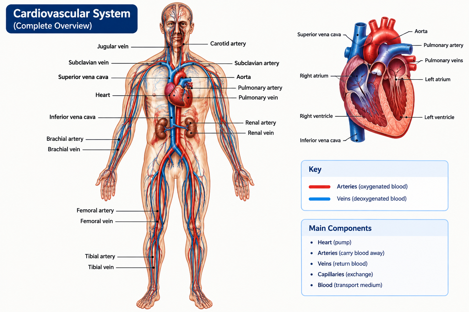
Major Components
Heart: muscular organ that pumps blood.
Arteries: carry blood away from the heart.
Veins: return blood toward the heart.
Capillaries: microscopic vessels where exchange occurs.
Blood: circulating connective tissue transported through the vessels.
Figure: Realistic labelled overview of the human cardiovascular system.
2. Parts of the Cardiovascular System
Component
Main structural features
Major function
Heart
Four chambers, valves, muscular wall and conducting system
Pumps blood through pulmonary and systemic circulation
Arteries
Thick muscular and elastic walls
Carry blood away from heart and distribute it to tissues
Arterioles
Small vessels with prominent smooth muscle
Regulate blood flow and vascular resistance
Capillaries
Single endothelial layer with basement membrane
Exchange of gases, nutrients and wastes
Venules
Small vessels draining capillary beds
Collect blood from capillaries
Veins
Thin walls and relatively large lumen
Return blood to heart and act as blood reservoirs
3. Gross Anatomy of the Heart
The heart is a hollow, muscular organ situated in the thoracic cavity
between the two lungs in the middle mediastinum. It is approximately
the size of a closed fist and is slightly conical in shape.
Location
Located in the middle mediastinum.
Situated between the right and left lungs.
Approximately two-thirds lies to the left of the median plane.
The apex is directed downward, forward and to the left.
The base is directed upward and posteriorly.
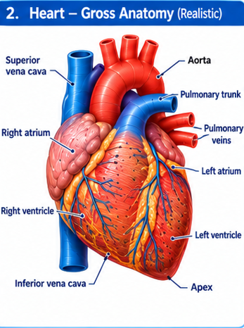
Figure: Gross anatomy of the human heart showing chambers,
major vessels, surfaces and apex.
External Features
The heart has an apex, base, diaphragmatic surface, sternocostal
surface and pulmonary surfaces. The coronary sulcus separates the
atria from the ventricles, while interventricular grooves mark the
position of the interventricular septum.
Major Blood Vessels Associated with Heart
Superior vena cava
Inferior vena cava
Pulmonary trunk
Right and left pulmonary arteries
Four pulmonary veins
Aorta
4. Microscopic Anatomy of the Heart
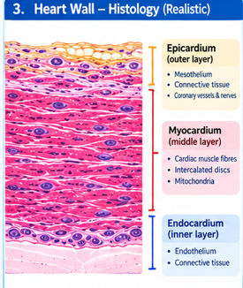
Histologically, the wall of the heart consists of three major layers:
endocardium, myocardium and epicardium.
Figure: Microscopic organization of the heart wall.
4.1 Endocardium
The endocardium is the innermost layer of the heart. It lines the
chambers and covers the cardiac valves.
Composed mainly of simple squamous endothelial cells.
Supported by connective tissue.
Contains small blood vessels, nerves and specialized conducting fibres in deeper regions.
Provides a smooth surface for blood flow.
4.2 Myocardium
The myocardium is the thick middle muscular layer and is the main
functional component responsible for contraction of the heart.
Composed of cardiac muscle fibres.
Fibres are branched and striated.
Usually contain one centrally located nucleus.
Cells are connected by intercalated discs.
Contains abundant mitochondria to support continuous contraction.
4.3 Epicardium
The epicardium forms the outer surface of the heart and corresponds
to the visceral layer of serous pericardium.
Covered by simple squamous mesothelium.
Contains connective tissue and variable amounts of adipose tissue.
Contains coronary blood vessels and nerves.
Exam point:
The myocardium is the thickest layer because it
performs the mechanical work of pumping blood. The left ventricular
myocardium is much thicker than the right ventricular myocardium
because it pumps blood through the high-resistance systemic circulation.
5. Chambers of the Heart
The heart is divided into four chambers: right atrium, right ventricle,
left atrium and left ventricle. The right and left sides are separated
by interatrial and interventricular septa.
Right Atrium
Receives deoxygenated blood from systemic circulation.
Receives blood through superior and inferior vena cava.
Receives blood from coronary sinus.
Communicates with right ventricle through tricuspid valve.
Right Ventricle
Receives blood from right atrium.
Pumps blood into pulmonary trunk.
Contains trabeculae carneae.
Contains papillary muscles and chordae tendineae.
Left Atrium
Receives oxygenated blood from four pulmonary veins.
Transfers blood to left ventricle.
Communicates through the mitral valve.
Left Ventricle
Receives oxygenated blood from left atrium.
Pumps blood into the aorta.
Has the thickest myocardium.
Generates high pressure for systemic circulation.
6. Cardiac Valves
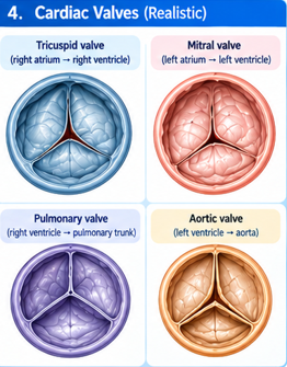
Cardiac valves maintain one-way movement of blood through the heart
and prevent backflow.
Valve
Location
Function
Tricuspid valve
Right atrium → right ventricle
Prevents backflow into right atrium during ventricular contraction
Mitral/Bicuspid valve
Left atrium → left ventricle
Prevents backflow into left atrium
Pulmonary semilunar valve
Right ventricle → pulmonary trunk
Prevents backflow from pulmonary trunk
Aortic semilunar valve
Left ventricle → aorta
Prevents backflow from aorta
Figure: Realistic labelled illustration of the four cardiac valves.
Chordae Tendineae and Papillary Muscles
The atrioventricular valves are attached to papillary muscles through
fibrous cords called chordae tendineae. During ventricular contraction,
papillary muscles contract and help prevent inversion of the valve
cusps into the atria.
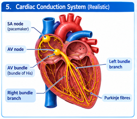
7. Conducting System of the Heart
The conducting system consists of specialized cardiac muscle cells
that generate and conduct electrical impulses responsible for
coordinated contraction of the heart.
Figure: Cardiac conducting system showing SA node, AV node,
AV bundle, bundle branches and Purkinje fibres.
Major Components
Sinoatrial node:
Located in the right atrium near the opening of the superior vena cava.
It acts as the normal pacemaker of the heart.
Atrioventricular node:
Located near the lower part of the interatrial septum. It delays
conduction so that the atria can complete contraction before the
ventricles contract.
Atrioventricular bundle:
Conducts impulses from the AV node into the interventricular septum.
Right and left bundle branches:
Conduct impulses toward the respective ventricles.
Purkinje fibres:
Distribute impulses throughout ventricular myocardium.
8. Coronary Circulation
The myocardium receives its blood supply through the coronary arteries.
These arteries arise from the ascending aorta immediately above the
aortic valve.
Right Coronary Artery
The right coronary artery supplies major portions of the right atrium,
right ventricle and parts of the conducting system. Important branches
include the right marginal artery and, in many individuals, the
posterior interventricular artery.
Left Coronary Artery
The left coronary artery divides mainly into the anterior
interventricular artery and circumflex artery. It supplies a large
portion of the left ventricle, interventricular septum and parts of
the left atrium.
Coronary Sinus
The coronary sinus collects most venous blood from the myocardium and
opens into the right atrium.
Important:
The coronary circulation is essential because cardiac muscle cannot
depend directly on blood present inside the cardiac chambers for its
oxygen requirements.
9. Gross and Microscopic Anatomy of Arteries
Arteries are blood vessels that carry blood away from the heart.
Most arteries carry oxygenated blood, although the pulmonary arteries
carry deoxygenated blood.
Three Basic Layers of Arterial Wall
Tunica intima:
Inner endothelial lining with supporting connective tissue.
Tunica media:
Middle layer containing smooth muscle and elastic fibres.
It is particularly well developed in arteries.
Tunica adventitia:
Outer connective tissue layer that supports and protects the vessel.
Types of Arteries
Type
Main feature
Example
Function
Elastic artery
Many elastic lamellae in tunica media
Aorta
Maintains continuous blood flow and dampens pressure fluctuations
Muscular artery
Prominent smooth muscle
Femoral artery
Distributes blood to organs
Arteriole
Small diameter with smooth muscle
Small tissue vessels
Controls peripheral resistance and tissue blood flow
Figure: Realistic microscopic illustration of an arterial wall.
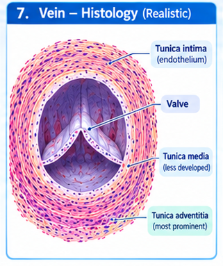
10. Gross and Microscopic Anatomy of Veins
Veins carry blood toward the heart. They generally have thinner walls
and larger lumina than corresponding arteries.
Structure
Tunica intima is relatively thin.
Tunica media contains less smooth muscle than arteries.
Tunica adventitia is often the thickest layer.
Many medium-sized veins contain valves.
Large veins may contain longitudinal smooth muscle in the adventitia.
Major Veins
Superior vena cava
Inferior vena cava
Pulmonary veins
Jugular veins
Subclavian veins
Renal veins
Femoral veins
Figure: Realistic microscopic structure of a vein.
11. Gross and Microscopic Anatomy of Capillaries
Capillaries are the smallest blood vessels and form networks between
arterioles and venules. Their extremely thin walls allow efficient
exchange between blood and tissues.
Microscopic Structure
Single layer of endothelial cells.
Basement membrane.
No well-developed tunica media.
Pericytes may be associated with the vessel wall.
Types of Capillaries
Type
Structure
Common location
Continuous
Continuous endothelial lining
Muscle, skin, brain
Fenestrated
Endothelial pores
Endocrine glands, intestinal villi, kidneys
Sinusoidal
Large irregular gaps and discontinuous basement membrane
Liver, spleen, bone marrow
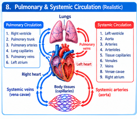
12. Pulmonary and Systemic Circulation
Pulmonary Circulation
Pulmonary circulation carries deoxygenated blood from the right ventricle
to the lungs and returns oxygenated blood to the left atrium.
Right ventricle → pulmonary trunk → pulmonary arteries →
pulmonary capillaries → pulmonary veins → left atrium.
Systemic Circulation
Systemic circulation carries oxygenated blood from the left ventricle
to body tissues and returns deoxygenated blood to the right atrium.
Regulate blood flow and systemic vascular resistance.
Capillaries
Exchange oxygen, carbon dioxide, nutrients, hormones and wastes.
Veins
Return blood to heart and serve as capacitance vessels.
Overall Functions
Transport of oxygen from lungs to tissues.
Transport of carbon dioxide from tissues to lungs.
Transport of nutrients from digestive tract.
Transport of hormones and other chemical messengers.
Removal of metabolic wastes.
Maintenance of body temperature.
Maintenance of acid-base and fluid balance.
Contribution to immune defense.
Maintenance of tissue perfusion and homeostasis.
14. Complete 10-Mark Long Questions
Important:
The following answers are designed to be directly prepared for
MSc II Semester MB 551 long-answer questions. In the examination,
use a clear heading structure and draw a labelled diagram wherever
appropriate.
LQ 1. Describe the gross and microscopic anatomy of the heart and explain its functions.
10 marks
Introduction
The heart is a hollow muscular organ of the cardiovascular system
that functions as a pump. It maintains continuous circulation of
blood through pulmonary and systemic circuits.
Location and General Structure
The heart is located in the middle mediastinum between the lungs.
It has an apex directed downward and to the left and a broad base
directed posteriorly. It is enclosed by the pericardium.
Chambers
Right atrium
Right ventricle
Left atrium
Left ventricle
Major Vessels
The superior and inferior venae cavae enter the right atrium.
The pulmonary trunk leaves the right ventricle. Four pulmonary
veins enter the left atrium, while the aorta leaves the left ventricle.
Microscopic Anatomy
The heart wall consists of endocardium, myocardium and epicardium.
The endocardium is lined by endothelium. The myocardium consists
of specialized cardiac muscle fibres with intercalated discs.
The epicardium consists of mesothelium and connective tissue
containing vessels, nerves and adipose tissue.
Valves
The tricuspid, mitral, pulmonary and aortic valves maintain
unidirectional blood flow and prevent backflow.
Functions
Maintains pulmonary circulation.
Maintains systemic circulation.
Generates blood pressure.
Delivers oxygen and nutrients to tissues indirectly through blood.
Removes carbon dioxide and metabolic wastes through circulation.
Supports hormonal and immune transport.
Conclusion
Thus, the structural organization of the heart is closely related
to its pumping function. The four chambers, valves, muscular wall
and conducting system work together to maintain effective circulation.
LQ 2. Describe the gross and microscopic anatomy of arteries, veins and capillaries. Add their functions.
10 marks
Introduction
Blood vessels form a continuous vascular network connecting the
heart with body tissues. They include arteries, arterioles,
capillaries, venules and veins.
Arteries
Arteries carry blood away from the heart. Their walls contain
three layers: tunica intima, tunica media and tunica adventitia.
The tunica media is particularly well developed.
Elastic arteries contain numerous elastic lamellae and help
maintain continuous blood flow. Muscular arteries contain
prominent smooth muscle and distribute blood to organs.
Veins
Veins return blood toward the heart. They have thinner walls,
larger lumina and less developed tunica media than arteries.
Many medium-sized veins possess valves that prevent backflow.
Capillaries
Capillaries consist primarily of a single layer of endothelial
cells supported by a basement membrane. Their thin walls permit
exchange of gases, nutrients, water and wastes.
Comparison
Feature
Artery
Vein
Capillary
Direction
Away from heart
Toward heart
Between arterioles and venules
Wall
Thick
Thin
Extremely thin
Muscular layer
Well developed
Less developed
Absent
Major function
Distribution
Return/storage
Exchange
Conclusion
The structure of each vessel is adapted to its function. Thick
muscular arterial walls tolerate and regulate pressure, veins
facilitate low-pressure return, and extremely thin capillaries
permit exchange with tissues.
LQ 3. Describe the chambers and valves of the heart and explain their functions.
10 marks
Four Chambers
The right atrium receives systemic venous blood and passes it to
the right ventricle through the tricuspid valve. The right ventricle
pumps blood into the pulmonary trunk.
The left atrium receives oxygenated blood from pulmonary veins and
passes it to the left ventricle through the mitral valve. The left
ventricle pumps blood into the aorta.
Valves
Tricuspid valve: between right atrium and right ventricle.
Mitral valve: between left atrium and left ventricle.
Pulmonary valve: between right ventricle and pulmonary trunk.
Aortic valve: between left ventricle and aorta.
Functional Importance
The valves open and close according to pressure differences.
Atrioventricular valves prevent ventricular blood from returning
to the atria during systole, while semilunar valves prevent blood
from returning from the great arteries into the ventricles during
diastole.
Therefore, coordinated chamber contraction and valve activity
maintain one-way circulation.
LQ 4. Describe the conducting system of the heart and explain its functional significance.
10 marks
Introduction
The conducting system is composed of specialized cardiac muscle
cells capable of generating and transmitting electrical impulses.
Components
Sinoatrial node
Atrioventricular node
Atrioventricular bundle
Right and left bundle branches
Purkinje fibres
Sequence of Impulse Conduction
The impulse normally begins in the sinoatrial node. It spreads
through the atrial myocardium and reaches the atrioventricular
node. The impulse is briefly delayed at the AV node before passing
through the AV bundle and bundle branches to Purkinje fibres.
Purkinje fibres distribute the impulse through the ventricular
myocardium, producing coordinated ventricular contraction.
Functions
Generates rhythmic cardiac impulses.
Coordinates atrial contraction.
Provides controlled delay before ventricular contraction.
Coordinates ventricular contraction.
Maintains effective cardiac pumping.
LQ 5. Explain pulmonary and systemic circulation with a suitable diagram.
10 marks
Pulmonary Circulation
Pulmonary circulation begins at the right ventricle. Deoxygenated
blood is pumped through the pulmonary trunk and pulmonary arteries
to the lungs. Gas exchange occurs in pulmonary capillaries.
Oxygenated blood returns through pulmonary veins to the left atrium.
Right ventricle → pulmonary trunk → pulmonary arteries →
lung capillaries → pulmonary veins → left atrium
Systemic Circulation
Systemic circulation begins at the left ventricle. Oxygenated
blood is pumped into the aorta and distributed through arteries
and arterioles to tissue capillaries. Deoxygenated blood returns
through venules and veins to the superior and inferior venae cavae.
Systemic circulation supplies tissues with oxygen and nutrients.
Both circuits work together to maintain continuous circulation.
LQ 6. Describe the coronary circulation and explain its importance.
10 marks
Coronary circulation supplies oxygenated blood to the myocardium
and removes metabolic wastes from cardiac muscle.
Arterial Supply
The right and left coronary arteries arise from the ascending aorta.
Their branches distribute blood to different regions of the heart.
Venous Drainage
Most cardiac venous blood drains into the coronary sinus, which
opens into the right atrium.
Importance
Provides oxygen to cardiac muscle.
Supplies nutrients to cardiomyocytes.
Removes carbon dioxide and metabolic waste.
Maintains continuous myocardial contraction.
Clinical correlation:
Obstruction of coronary blood flow can reduce oxygen delivery
to cardiac muscle and may cause myocardial ischemia or infarction.
LQ 7. Describe the microscopic structure of the heart wall and correlate it with its functions.
10 marks
Endocardium
The endocardium forms the inner lining of the cardiac chambers.
Its endothelial surface provides a smooth blood-contacting surface.
Myocardium
The myocardium is composed of cardiac muscle cells. These cells
are striated, branched and connected by intercalated discs.
Gap junctions facilitate electrical communication between cells,
while mechanical junctions provide strong attachment.
Epicardium
The epicardium covers the external surface of the heart. It
contains connective tissue, adipose tissue, coronary vessels
and nerves and is covered by mesothelium.
Functional Correlation
Layer
Structural feature
Functional importance
Endocardium
Endothelium
Smooth internal surface and blood compatibility
Myocardium
Cardiac muscle
Contraction and pumping
Epicardium
Mesothelium and connective tissue
Protection and support of coronary structures
15. Separate 5-Mark Preparation
For 5 marks:
Start with a definition/introduction, give 4–6 organized points,
and finish with the main function or significance.
1. Gross Anatomy of Heart
The heart is a hollow muscular organ situated in the middle
mediastinum. It has four chambers: right atrium, right ventricle,
left atrium and left ventricle. The major vessels include venae
cavae, pulmonary trunk, pulmonary veins and aorta. The heart has
an apex and base and is surrounded by the pericardium.
Prevent backflow and maintain one-way circulation.
4. Conducting System
The conducting system includes SA node, AV node, AV bundle,
bundle branches and Purkinje fibres. It generates and distributes
electrical impulses and coordinates atrial and ventricular contraction.
5. Histology of Artery
Arteries have tunica intima, tunica media and tunica adventitia.
The tunica media is well developed and contains smooth muscle and
elastic fibres. Arteries withstand and regulate relatively high
blood pressure.
6. Histology of Vein
Veins have a relatively thin wall and large lumen. The tunica media
is less developed while the adventitia is prominent. Many veins
possess valves. Their major function is returning blood to the heart.
7. Capillaries
Capillaries consist mainly of endothelial cells and a basement
membrane. Their thin walls allow exchange of oxygen, carbon dioxide,
nutrients, water and wastes between blood and tissues.
8. Pulmonary Circulation
Deoxygenated blood flows from the right ventricle through the
pulmonary arteries to lung capillaries. After gas exchange,
oxygenated blood returns through pulmonary veins to the left atrium.
9. Systemic Circulation
Oxygenated blood leaves the left ventricle through the aorta and
travels through arteries, arterioles and tissue capillaries.
Deoxygenated blood returns through veins to the venae cavae and
enters the right atrium.
10. Coronary Circulation
Coronary arteries arise from the ascending aorta and supply the
myocardium. Cardiac venous blood drains mainly through the coronary
sinus into the right atrium. Coronary circulation is essential for
oxygen and nutrient supply to cardiac muscle.
11. Functions of Cardiovascular System
Transport of oxygen and carbon dioxide.
Transport of nutrients and hormones.
Removal of metabolic wastes.
Regulation of temperature and fluid balance.
Maintenance of tissue perfusion and homeostasis.
16. Separate 2-Mark Preparation
Question
Short Answer
What is the cardiovascular system?
A transport system consisting mainly of the heart, blood vessels
and blood that maintains circulation throughout the body.
Name the chambers of heart.
Right atrium, right ventricle, left atrium and left ventricle.
Name the layers of heart wall.
Endocardium, myocardium and epicardium.
Which is the thickest chamber of heart?
Left ventricle.
Name the four cardiac valves.
Tricuspid, mitral, pulmonary and aortic valves.
What is the function of cardiac valves?
They prevent backflow and maintain unidirectional blood flow.
What is SA node?
The sinoatrial node is the normal pacemaker of the heart that
initiates cardiac electrical impulses.
What is AV node?
A specialized conducting structure that delays and transmits
electrical impulses from atria toward ventricles.
Name the three layers of blood vessel wall.
Tunica intima, tunica media and tunica adventitia.
What are arteries?
Blood vessels that carry blood away from the heart.
What are veins?
Blood vessels that return blood toward the heart.
What are capillaries?
Microscopic vessels consisting mainly of a single endothelial
layer where exchange occurs between blood and tissues.
Name the types of capillaries.
Continuous, fenestrated and sinusoidal capillaries.
What is pulmonary circulation?
Circulation of blood between the heart and lungs for gas exchange.
What is systemic circulation?
Circulation of blood between the heart and body tissues.
What is coronary circulation?
Blood circulation supplying the myocardium through coronary vessels.
Why is the left ventricular wall thick?
Because it generates high pressure required to pump blood through
systemic circulation.
What is myocardium?
The thick cardiac muscle layer of the heart responsible for contraction.
What are Purkinje fibres?
Specialized conducting fibres that distribute electrical impulses
throughout the ventricular myocardium.
What is the function of capillaries?
Exchange of gases, nutrients, water, hormones and metabolic wastes.
SA node → AV node → AV bundle →
Bundle branches → Purkinje fibres
Important Exam Points
In a 10-mark answer, never write only the names of heart chambers.
Explain their structure and function.
Always mention the three layers of the heart wall:
endocardium, myocardium and epicardium.
For blood vessels, remember the three layers:
tunica intima, tunica media and tunica adventitia.
Explain why the left ventricular wall is thicker than the right
ventricular wall.
In histology questions, mention the cellular composition and
correlate it with function.
Draw a labelled heart diagram in a 10-mark answer whenever possible.
For conducting-system questions, write the complete sequence:
SA node → AV node → AV bundle → bundle branches → Purkinje fibres.
Clearly distinguish pulmonary circulation from systemic circulation.
Coronary circulation is important because the myocardium requires
its own blood supply for continuous contraction.
For artery, vein and capillary questions, include a comparison table
because it makes the structural differences easy to score.
Diagrams You Should Practice for LQ
1. Gross Heart
Label right atrium, right ventricle, left atrium, left ventricle,
aorta, pulmonary trunk, venae cavae and pulmonary veins.
2. Heart Wall
Label endocardium, myocardium, epicardium and pericardial region.
3. Cardiac Valves
Label tricuspid, mitral, pulmonary and aortic valves.
4. Conducting System
Label SA node, AV node, AV bundle, bundle branches and Purkinje fibres.
5. Blood Vessel Histology
Show tunica intima, tunica media and tunica adventitia.
6. Pulmonary + Systemic Circulation
Show the complete pathway from right heart to lungs to left heart
and then through systemic tissues.
Selected Clinical Correlations
Coronary Artery Disease
Narrowing or obstruction of coronary arteries can reduce blood supply
to the myocardium. Reduced myocardial oxygen delivery may cause
ischemia and, if prolonged and severe, myocardial infarction.
Valvular Disorders
Abnormal narrowing or incomplete closure of a valve can interfere
with normal one-way blood flow and alter cardiac workload.
Hypertension
Persistent elevation of arterial pressure increases the workload
imposed on the cardiovascular system and may contribute to structural
and functional changes in the heart and blood vessels.
How to Write a 10-Mark Cardiovascular LQ
Recommended Answer Pattern
Introduction / definition
Location and general description
Gross anatomy
Microscopic anatomy / histology
Functions of each major part
Relevant physiological significance
Simple labelled diagram
Clinical correlation if relevant
Short conclusion
This structure helps keep the answer organized and ensures that both
components of the MB 551 syllabus — anatomical structure and
functions — are covered.