MB 551 Human Anatomy & Physiology • 4 Hours

Urinary System

Detailed MSc Medical Microbiology / TU-style study notes covering the organs, gross anatomy, microscopic anatomy, functions, nephron structure, urine formation, renal regulation and composition of urine.

High-Weight Long Question Chapter: The urinary system can generate long questions from both anatomy and physiology. Prepare the kidney and nephron thoroughly, especially glomerular filtration, tubular reabsorption, tubular secretion, concentration of urine, hormonal regulation and composition of urine.

1. Introduction to the Urinary System

Basic organization and importance of the urinary system

Definition: The urinary system is an organ system responsible mainly for the formation and excretion of urine. It maintains the internal chemical environment of the body by regulating water balance, electrolyte concentration, acid-base balance, blood volume and blood pressure. It also removes metabolic waste products and contributes to endocrine functions such as erythropoietin production and activation of vitamin D.

The urinary system is closely associated with homeostasis. Although the kidneys are commonly described as organs that simply remove waste from the blood, their physiological role is much broader. The kidneys continuously filter plasma, selectively return useful substances to the blood, secrete additional substances into the tubular fluid and finally excrete the remaining material as urine.

The major organs of the urinary system are the two kidneys, two ureters, urinary bladder and urethra. The kidneys are the functional organs of the system because urine is formed within them. The ureters transport urine from the kidneys to the urinary bladder, the bladder temporarily stores urine and the urethra conducts urine from the bladder to the exterior.

Kidneys

Form urine, regulate body fluid composition, maintain acid-base balance and perform important endocrine functions.

Ureters

Muscular tubes that transport urine from the renal pelvis to the urinary bladder by peristaltic contractions.

Urinary Bladder

Hollow muscular organ that temporarily stores urine before micturition.

Urethra

Conducts urine from the urinary bladder to the exterior during micturition.

2. Different Organs of the Urinary System

Major organs and their basic functions

Organ Number Main function
Kidneys 2 Formation of urine, removal of metabolic wastes, regulation of water, electrolytes, pH and blood pressure; endocrine functions.
Ureters 2 Transport urine from renal pelvis to urinary bladder.
Urinary bladder 1 Temporary storage of urine and controlled emptying during micturition.
Urethra 1 Conducts urine from urinary bladder to the external environment.
Exam point: In a long answer, begin with the four organs of the urinary system, then discuss the kidney in greatest detail because the kidney is the principal organ responsible for urine formation.

3. Gross Anatomy of the Kidney

External and internal structure of the kidney

Realistic textbook-style gross anatomy of the human kidney showing cortex, medulla, renal pyramids, calyces and renal pelvis
Figure: Gross structure of the kidney. Add the corresponding realistic textbook-style anatomical image to the images folder.

The kidneys are paired, bean-shaped, highly vascular organs located on the posterior abdominal wall. They are retroperitoneal organs, meaning that they lie behind the peritoneum. They are positioned on either side of the vertebral column. The right kidney usually lies slightly lower than the left kidney because of the presence of the liver above it.

Each adult kidney is approximately 10–12 cm long, 5–7 cm wide and about 3 cm thick, although the exact dimensions vary between individuals. The medial border is concave and contains the renal hilum, through which the renal artery, renal vein, nerves, lymphatics and ureter enter or leave the kidney.

External Features

  • Superior pole
  • Inferior pole
  • Anterior surface
  • Posterior surface
  • Lateral convex border
  • Medial concave border
  • Renal hilum

Coverings of Kidney

The kidney is protected by several layers. From superficial to deep, the important coverings include the renal fascia, perirenal fat, fibrous renal capsule and the underlying renal tissue.

Covering Importance
Renal fascia Anchors the kidney to surrounding structures and helps maintain its position.
Perirenal fat Provides cushioning and protection.
Fibrous renal capsule Thin connective tissue layer directly surrounding the kidney.

Internal Structure of Kidney

On section, the kidney can be divided into an outer renal cortex and an inner renal medulla. The cortex extends inward between the medullary pyramids as renal columns.

The medulla consists mainly of several cone-shaped renal pyramids. The apex of each pyramid is called the renal papilla. Urine passes from the papilla into a minor calyx, then into major calyces and finally into the renal pelvis.

Collecting ducts
→
Renal papilla
→
Minor calyx
→
Major calyx
→
Renal pelvis
→
Ureter

4. Microscopic Anatomy of the Kidney

Histological organization of renal tissue

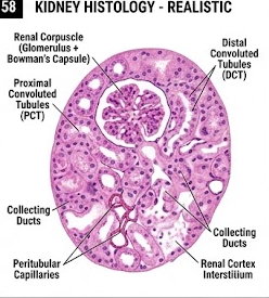
Realistic kidney histology showing renal corpuscles, proximal tubules, distal tubules and collecting ducts
Figure: Microscopic organization of kidney showing renal corpuscles and renal tubules.

The functional microscopic organization of the kidney is based on the nephron and collecting duct system. The renal cortex contains renal corpuscles, proximal convoluted tubules and distal convoluted tubules. The medulla contains loops of Henle and collecting ducts, together with associated blood vessels.

The cortex has a granular appearance because it contains numerous renal corpuscles and convoluted tubules. The medulla has a more striated appearance because of the parallel arrangement of loops of Henle, collecting ducts and blood vessels.

Renal Corpuscle

The renal corpuscle is the initial component of the nephron and is responsible for the first step in urine formation: glomerular filtration. It consists of a glomerulus surrounded by Bowman's capsule.

The glomerulus is a tuft of fenestrated blood capillaries supplied by the afferent arteriole and drained by the efferent arteriole. Bowman's capsule has a parietal layer and a visceral layer. The visceral layer is formed by specialized cells called podocytes.

Filtration Barrier

The glomerular filtration barrier is highly specialized. It prevents most blood cells and large plasma proteins from passing into the filtrate while allowing water and many small dissolved substances to pass.

  1. Fenestrated glomerular endothelium
  2. Glomerular basement membrane
  3. Filtration slits between podocyte foot processes

Proximal Convoluted Tubule

The proximal convoluted tubule is lined mainly by simple cuboidal epithelial cells with a prominent brush border formed by numerous microvilli. These microvilli greatly increase the surface area available for reabsorption.

The proximal tubule performs extensive reabsorption. Under normal conditions, a large proportion of filtered sodium, water, glucose, amino acids and bicarbonate is reabsorbed here. It also participates in secretion of hydrogen ions, ammonium and several organic substances.

Loop of Henle

The loop of Henle extends from the proximal tubule toward the renal medulla and then returns toward the cortex. It has descending and ascending limbs with different permeability characteristics.

The loop of Henle is essential for establishing the medullary osmotic gradient that allows the kidneys to produce concentrated urine when water conservation is necessary.

Distal Convoluted Tubule

The distal convoluted tubule is lined by simple cuboidal epithelium but has fewer and shorter microvilli than the proximal tubule. It participates in regulation of sodium, potassium, calcium and acid-base balance.

Collecting Duct

Collecting ducts receive tubular fluid from several nephrons and carry it through the cortex and medulla toward the renal papilla. Their water permeability is strongly regulated by antidiuretic hormone (ADH).

In the presence of ADH, collecting ducts become more permeable to water, allowing water to move into the hyperosmotic renal medulla. Consequently, more concentrated urine is produced.

5. Nephron: Structural and Functional Unit of Kidney

Realistic textbook-style nephron showing renal corpuscle, proximal tubule, loop of Henle, distal tubule and collecting duct
Figure: Detailed structure of a nephron and associated collecting duct.
Nephron: The nephron is the structural and functional unit of the kidney. Each kidney contains approximately one million nephrons, although the exact number varies.

Each nephron begins with a renal corpuscle and continues through a sequence of renal tubules. Its major components are the renal corpuscle, proximal convoluted tubule, loop of Henle, distal convoluted tubule and connecting segment.

The tubular system is closely associated with blood vessels. This arrangement is essential because filtration occurs from glomerular capillaries into Bowman's space, while reabsorption and secretion occur between tubular fluid and the surrounding peritubular circulation.

Part Main structural feature Main physiological role
Renal corpuscle Glomerulus + Bowman's capsule Glomerular filtration
Proximal tubule Simple cuboidal cells with brush border Bulk reabsorption and secretion
Loop of Henle Descending and ascending limbs Medullary osmotic gradient and urine concentration
Distal tubule Simple cuboidal epithelium Fine regulation of electrolytes and pH
Collecting duct Distinct epithelial cells Final water and electrolyte regulation

Types of Nephrons

Cortical Nephrons

Their renal corpuscles are located mainly in the outer cortex. Their loops of Henle are relatively short and extend only a limited distance into the medulla.

Juxtamedullary Nephrons

Their renal corpuscles lie close to the corticomedullary junction. They possess long loops of Henle and are particularly important in generating concentrated urine.

6. Renal Blood Circulation

Realistic renal circulation showing renal artery, afferent arteriole, glomerulus, efferent arteriole and peritubular capillaries
Figure: Renal circulation associated with filtration and tubular exchange.

The kidneys receive a very large blood supply because they must continuously filter plasma and regulate its composition. Blood enters the kidney through the renal artery, which divides into progressively smaller arteries and eventually supplies the glomeruli through afferent arterioles.

Renal artery
→
Afferent arteriole
→
Glomerulus
→
Efferent arteriole
→
Peritubular capillaries / Vasa recta

The unusual arrangement of two capillary beds in series is important for renal function. The first capillary bed is the glomerulus, where high pressure promotes filtration. The second is the peritubular capillary network, which supports reabsorption and secretion.

In juxtamedullary nephrons, the efferent arterioles form long, straight vessels called the vasa recta. These vessels participate in maintaining the osmotic gradient of the renal medulla.

7. Ureter

Gross and microscopic anatomy with function

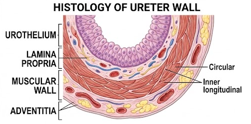
Realistic histological structure of ureter showing urothelium, muscular wall and connective tissue
Figure: Microscopic structure of the ureter.

Each ureter is a muscular tube that carries urine from the renal pelvis to the urinary bladder. It is approximately 25–30 cm long in adults, although individual measurements vary.

Microscopic Structure

The ureter has three major layers:

  1. Mucosa: lined by transitional epithelium (urothelium), which allows considerable distension.
  2. Muscularis: mainly smooth muscle arranged in longitudinal and circular patterns. The lower portion also has an additional outer longitudinal layer.
  3. Adventitia: outer connective tissue containing blood vessels, lymphatics and nerves.

Urine is transported through the ureter mainly by rhythmic peristaltic contractions of its smooth muscle. Therefore, urine does not depend simply on gravity to reach the urinary bladder.

8. Urinary Bladder

Gross and microscopic structure

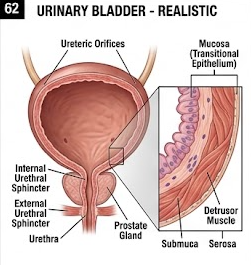
Realistic gross anatomy of urinary bladder showing ureters, trigone and urethra
Figure: Gross structure of urinary bladder.

The urinary bladder is a hollow muscular organ located in the pelvis. Its principal function is temporary storage of urine until it is expelled during micturition.

When empty, the bladder is relatively collapsed. As urine enters, the bladder expands because its wall can stretch while maintaining relatively low pressure over a considerable volume.

Important Parts

  • Apex
  • Body
  • Fundus
  • Neck
  • Trigone

Microscopic Structure

The mucosa is lined by transitional epithelium, also called urothelium. Beneath it is connective tissue, followed by a thick muscular layer called the detrusor muscle.

The transitional epithelium is particularly suited to the urinary bladder because its superficial cells can change shape as the bladder distends.

Important: The detrusor muscle contracts during bladder emptying. The internal urethral sphincter is smooth muscle, while the external urethral sphincter is skeletal muscle and is under voluntary control.

9. Urethra

The urethra is the terminal passage of the urinary system. It carries urine from the urinary bladder to the exterior.

The female urethra is shorter than the male urethra. The male urethra is longer and is divided anatomically into prostatic, membranous and spongy portions. In males, it serves as a common passage for both urinary and reproductive systems.

Microscopic Structure

The epithelial lining varies along the length of the urethra. It may include transitional epithelium near the bladder, followed by pseudostratified or stratified columnar epithelium in portions, and stratified squamous epithelium near the external opening.

10. Functions of the Kidneys

1. Excretion

Removes urea, uric acid, creatinine, drug metabolites and other unwanted substances from the body.

2. Water Balance

Regulates water excretion according to the body's hydration status.

3. Electrolyte Balance

Regulates sodium, potassium, calcium, phosphate and other ions.

4. Acid-Base Balance

Excretes hydrogen ions and regulates bicarbonate handling.

5. Blood Pressure

Influences blood pressure through sodium-water balance and the renin-angiotensin-aldosterone system.

6. Endocrine Function

Produces erythropoietin and renin and activates vitamin D to calcitriol.

11. Mechanism of Urine Formation

Major physiological processes responsible for urine production

Realistic renal physiology diagram showing glomerular filtration, tubular reabsorption and tubular secretion
Figure: Major steps involved in urine formation.
Urine formation is a highly regulated process by which the kidneys filter blood plasma, selectively reabsorb useful substances, secrete additional substances into the tubular fluid and excrete the final urine.

Urine formation can be understood through three fundamental processes:

Glomerular filtration
+
Tubular reabsorption
+
Tubular secretion
→
Urinary excretion
Urinary excretion = Filtration − Reabsorption + Secretion

This relationship is central to understanding renal physiology. A substance may be filtered, partially or completely reabsorbed, secreted, or both reabsorbed and secreted. The final amount appearing in urine depends on the balance among these processes.

12. Glomerular Filtration

Glomerular filtration is the first step in urine formation. It occurs in the renal corpuscle when fluid and small dissolved substances move from the glomerular capillaries into Bowman's space.

Filtration is driven mainly by the pressure generated within the glomerular capillaries. The filtration barrier allows water and small solutes to pass while restricting blood cells and most plasma proteins.

Forces Determining Filtration

Glomerular filtration is governed by Starling forces. The main filtration pressure is the glomerular capillary hydrostatic pressure. Opposing forces include plasma colloid osmotic pressure and the hydrostatic pressure within Bowman's space.

Net Filtration Pressure = Glomerular Capillary Hydrostatic Pressure − (Bowman's Space Hydrostatic Pressure + Plasma Colloid Osmotic Pressure)

The resulting filtrate is similar to plasma in its concentration of many small molecules, but it normally contains very little protein and essentially no blood cells.

Glomerular Filtration Rate

The glomerular filtration rate (GFR) is the volume of filtrate formed by both kidneys per unit time. In a healthy young adult, a commonly cited value is approximately 125 mL/min, although normal values vary with age, body size and sex.

Exam point: GFR is an important indicator of renal function. A significant persistent reduction in GFR may indicate impaired kidney function.

13. Tubular Reabsorption

Selective recovery of useful substances from tubular fluid

Tubular reabsorption is the process by which substances present in the tubular fluid move back into the blood. It is essential because glomerular filtration is relatively non-selective for many small molecules.

Without extensive reabsorption, enormous quantities of water, glucose, amino acids, sodium and other useful substances would be lost in urine.

Reabsorption may occur through the epithelial cells (transcellular pathway) or between adjacent cells (paracellular pathway). Transport may be passive or active and is regulated according to the body's needs.

Reabsorption Along Different Tubular Segments

Segment Important reabsorptive processes
Proximal tubule Major reabsorption of sodium and water; almost complete reabsorption of filtered glucose and amino acids under normal conditions; substantial bicarbonate reabsorption.
Descending limb Highly permeable to water; water leaves the tubule into the hyperosmotic medulla.
Ascending limb Relatively impermeable to water and important for NaCl reabsorption.
Distal tubule Fine regulation of sodium, chloride, calcium and acid-base balance.
Collecting duct Variable water reabsorption under the influence of ADH; also contributes to potassium and acid-base regulation.

Glucose Reabsorption

Normally, virtually all filtered glucose is reabsorbed in the proximal tubule. Glucose enters proximal tubular cells through sodium-glucose cotransport mechanisms and then moves into the interstitial fluid and blood. When plasma glucose becomes sufficiently high, the transport maximum may be exceeded and glucose appears in urine, producing glycosuria.

14. Tubular Secretion

Tubular secretion is the transfer of selected substances from the blood into the tubular fluid. It complements filtration and is particularly important for substances that are poorly filtered or require additional elimination.

Important substances secreted by renal tubules include hydrogen ions, potassium ions, ammonium and various organic acids and bases. Many drugs and drug metabolites are also secreted by the proximal tubule.

Tubular secretion has major roles in potassium regulation and acid-base balance. For example, secretion of hydrogen ions contributes to removal of acid from the body, while ammonium excretion allows the kidney to eliminate acid without producing an excessively low urine pH.

15. Concentration and Dilution of Urine

Countercurrent mechanism and renal medullary osmotic gradient

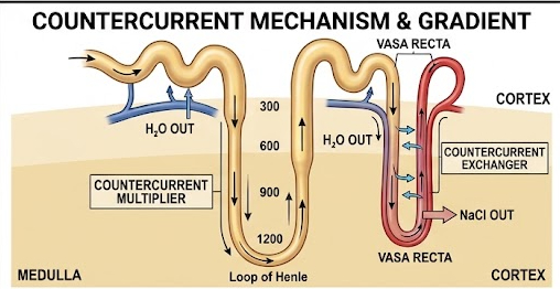
Realistic renal countercurrent mechanism showing loop of Henle and vasa recta
Figure: Countercurrent mechanism involved in concentration of urine.

One of the remarkable functions of the kidney is its ability to produce either dilute urine when excess water must be removed or concentrated urine when water must be conserved.

The ability to concentrate urine depends mainly on the countercurrent mechanism involving the loop of Henle, the collecting ducts and the vasa recta.

Role of Descending Limb

The descending limb of the loop of Henle is relatively permeable to water. As tubular fluid moves deeper into the increasingly hyperosmotic medulla, water leaves the tubule by osmosis. Consequently, the tubular fluid becomes more concentrated.

Role of Ascending Limb

The ascending limb is relatively impermeable to water. Sodium and chloride are transported out of the tubular fluid, particularly in the thick ascending limb. This contributes to the high osmolarity of the renal medulla while the tubular fluid becomes progressively diluted.

Role of Vasa Recta

The vasa recta are specialized capillaries associated with juxtamedullary nephrons. Their hairpin arrangement and slow blood flow allow them to function as a countercurrent exchanger and help prevent washout of the medullary osmotic gradient.

Role of ADH

Antidiuretic hormone increases the water permeability of the collecting ducts by promoting insertion of aquaporin-2 water channels into the apical membrane of principal cells. Water can therefore be reabsorbed into the hyperosmotic medullary interstitium, producing concentrated urine.

16. Hormonal Regulation of Renal Function

Hormone Main renal effect
ADH Increases water reabsorption in collecting ducts and promotes concentration of urine.
Aldosterone Increases sodium reabsorption and potassium secretion mainly in the distal nephron.
Angiotensin II Promotes sodium retention, stimulates aldosterone and ADH release, and contributes to maintenance of blood pressure.
Renin Initiates the renin-angiotensin-aldosterone system in response to appropriate renal stimuli such as reduced renal perfusion.
Atrial natriuretic peptide Promotes sodium excretion and opposes excessive volume expansion.
Parathyroid hormone Increases renal calcium reabsorption and decreases phosphate reabsorption in the proximal tubule.

17. Juxtaglomerular Apparatus

The juxtaglomerular apparatus (JGA) is a specialized region where the distal tubule comes into close association with the vascular pole of the renal corpuscle. It plays an important role in regulation of GFR and systemic blood pressure.

The main components are:

  1. Macula densa: specialized cells of the distal tubule that monitor tubular sodium chloride concentration.
  2. Juxtaglomerular cells: modified smooth muscle cells in the afferent arteriole that produce and release renin.
  3. Extraglomerular mesangial cells: cells involved in communication and support within the apparatus.

The JGA participates in tubuloglomerular feedback and in the renin-angiotensin-aldosterone system.

18. Role of Kidney in Acid-Base Balance

The kidneys play a major role in maintaining the pH of body fluids. They act more slowly than respiratory mechanisms but provide powerful long-term regulation of acid-base balance.

The kidneys maintain acid-base balance by:

  • Reabsorbing filtered bicarbonate.
  • Secreting hydrogen ions into tubular fluid.
  • Producing and excreting ammonium.
  • Generating new bicarbonate during net acid excretion.

In the proximal tubule, bicarbonate is extensively reclaimed. Hydrogen ion secretion and carbonic anhydrase-dependent reactions facilitate this process.

During chronic acid loading, renal ammonium production and acid excretion can increase substantially, helping restore systemic acid-base balance.

19. Micturition

Micturition is the process of emptying urine from the urinary bladder through the urethra.

As urine accumulates in the bladder, stretch receptors in the bladder wall are activated. Sensory information travels to the spinal cord, particularly the sacral micturition centers. The process is modified by higher centers of the brain, allowing voluntary control over external sphincter activity.

Basic Events

  1. Urine enters the bladder.
  2. Bladder wall stretches.
  3. Stretch receptors are activated.
  4. Sacral parasympathetic pathways are activated.
  5. Detrusor muscle contracts.
  6. Internal urethral outlet resistance decreases.
  7. External urethral sphincter is voluntarily relaxed.
  8. Urine passes through the urethra.

20. Composition of Normal Urine

Normal urine is a clear, usually pale-yellow fluid produced by the kidneys. Its exact composition changes according to water intake, diet, metabolic activity, hormones, exercise and other physiological conditions.

Water forms the major component of urine, while the remaining fraction consists of dissolved organic and inorganic substances.

Component Normal significance
Water Major component and medium for dissolved wastes.
Urea Major nitrogenous waste derived mainly from protein metabolism.
Creatinine Waste product associated with muscle creatine metabolism.
Uric acid End product of purine metabolism.
Electrolytes Include sodium, potassium, chloride, phosphate, sulfate and other ions in variable concentrations.
Ammonium Important in renal acid excretion.
Pigments Mainly urochrome-related pigments contribute to the yellow color.

Substances Normally Absent or Present Only in Trace Amounts

  • Significant amounts of glucose
  • Significant amounts of protein
  • Blood cells
  • Large quantities of ketone bodies
  • Hemoglobin
  • Significant bilirubin
Clinical significance: The appearance of abnormal substances in urine may provide important information about disease. Examples include proteinuria, glycosuria, hematuria, ketonuria and bilirubinuria.

21. Physical Characteristics of Urine

Feature Normal description
Appearance Usually clear to slightly yellow.
Color Usually pale yellow to deeper yellow depending on concentration.
Odor Characteristic odor; may change after standing.
pH Variable, commonly mildly acidic but influenced by diet and metabolism.
Specific gravity Variable depending on concentration of dissolved substances and hydration status.
Daily volume Commonly around 1–2 L/day in adults, but strongly influenced by fluid intake and physiological conditions.

22. Clinical Correlations

Important clinical points for examination

Glomerulonephritis

Inflammation affecting glomerular structures can impair filtration and may result in proteinuria or hematuria.

Nephrotic Syndrome

Characterized by marked protein loss in urine and associated abnormalities such as hypoalbuminemia and edema.

Acute Kidney Injury

A sudden decline in renal function causing impaired excretion and disturbances of fluid, electrolyte and acid-base balance.

Chronic Kidney Disease

Persistent loss of kidney function that may progressively reduce filtration and disturb homeostasis.

Renal Calculi

Crystalline stones may develop in the urinary tract and cause severe pain or urinary obstruction.

Urinary Tract Infection

Infection may involve the urethra, bladder, ureters or kidneys, with bacteria being common causative agents.

Diabetes Mellitus

Hyperglycemia may exceed the renal glucose transport capacity, resulting in glycosuria and osmotic diuresis.

Diabetes Insipidus

Reduced ADH activity or renal response to ADH can impair water conservation and produce large volumes of dilute urine.

23. 10-Mark Long Questions

Complete written answers for major examination areas

LQ 1. Describe the different organs of the urinary system and discuss the gross and microscopic anatomy of the kidney.

Answer

The urinary system consists of two kidneys, two ureters, the urinary bladder and the urethra. It is responsible for urine formation, transport, storage and elimination. The kidneys are the major functional organs because they regulate the composition of body fluids and eliminate metabolic wastes.

Kidneys

The kidneys are paired retroperitoneal organs located on the posterior abdominal wall. They are bean-shaped and have a convex lateral border and a concave medial border. The medial border contains the renal hilum, through which the renal vessels, nerves and ureter pass.

Externally, the kidney is covered by a fibrous capsule and supported by surrounding connective tissue and fat. Internally, it is divided into an outer cortex and inner medulla. The medulla contains renal pyramids whose papillae drain urine into minor calyces. Minor calyces unite to form major calyces, which drain into the renal pelvis and subsequently into the ureter.

Microscopic Anatomy

Microscopically, the kidney consists primarily of nephrons and collecting ducts. Each nephron begins with a renal corpuscle consisting of a glomerulus enclosed by Bowman's capsule.

The proximal convoluted tubule is lined by cuboidal cells with a prominent brush border and performs extensive reabsorption. The loop of Henle consists of descending and ascending portions and contributes to the countercurrent mechanism. The distal convoluted tubule is involved in fine regulation of electrolytes and acid-base balance. Collecting ducts transport tubular fluid toward the renal papilla and regulate final water excretion under the influence of ADH.

Other Organs

The ureters are muscular tubes lined by transitional epithelium and transport urine by peristalsis. The urinary bladder is a muscular storage organ lined by urothelium and contains the detrusor muscle. The urethra conducts urine from the bladder to the exterior.

Therefore, the urinary system has a continuous structural arrangement from the microscopic nephron to the ureter, bladder and urethra, allowing urine to be formed, transported, stored and finally eliminated.

LQ 2. Describe the nephron with its microscopic structure and explain the functions of its different parts.

Answer

The nephron is the structural and functional unit of the kidney. Each nephron is specialized to filter plasma, reabsorb useful substances, secrete selected substances and form urine.

The nephron begins with the renal corpuscle and continues through the proximal tubule, loop of Henle and distal tubule. The tubular system is closely associated with a specialized vascular network.

1. Renal Corpuscle

The renal corpuscle contains a glomerular capillary tuft surrounded by Bowman's capsule. The glomerulus receives blood through the afferent arteriole and drains through the efferent arteriole. Filtration occurs across the glomerular filtration barrier.

2. Proximal Convoluted Tubule

The proximal tubule has cuboidal epithelial cells with abundant microvilli. It performs bulk reabsorption of sodium, water, bicarbonate, glucose and amino acids. It also secretes hydrogen ions, ammonium and various organic substances.

3. Loop of Henle

The loop of Henle extends into the medulla. The descending limb is highly permeable to water, whereas the ascending limb is relatively impermeable to water and actively contributes to sodium chloride transport. This difference is essential for the countercurrent mechanism.

4. Distal Convoluted Tubule

The distal tubule participates in regulation of sodium, potassium, calcium and acid-base balance. It is also closely associated with the macula densa of the juxtaglomerular apparatus.

5. Collecting Duct

Collecting ducts receive fluid from multiple nephrons and pass through the renal medulla. Their permeability to water is regulated by ADH, allowing the kidney to produce concentrated urine when necessary.

Thus, different parts of the nephron perform specialized but coordinated functions that collectively maintain the internal environment of the body.

LQ 3. Explain the mechanism of urine formation in detail.

Answer

Urine formation is a complex physiological process occurring in the nephrons of the kidneys. It involves selective filtration of plasma, recovery of useful substances and elimination of unwanted substances. The three major processes are glomerular filtration, tubular reabsorption and tubular secretion.

1. Glomerular Filtration

Blood enters the glomerulus through the afferent arteriole. Because glomerular capillary hydrostatic pressure is relatively high, water and small dissolved substances pass through the filtration barrier into Bowman's space.

The filtration barrier consists of fenestrated endothelium, glomerular basement membrane and podocyte filtration slits. Blood cells and most plasma proteins are retained within the circulation.

2. Tubular Reabsorption

The filtrate contains many substances that the body needs. Therefore, the renal tubules selectively return useful substances to the blood. The proximal tubule performs the largest amount of reabsorption. Sodium and water are extensively reabsorbed, while filtered glucose and amino acids are normally almost completely recovered.

The loop of Henle establishes the medullary concentration gradient. The descending limb permits water movement, whereas the ascending limb reabsorbs sodium chloride and is relatively impermeable to water.

The distal nephron and collecting ducts provide further fine regulation of sodium, potassium, water and acid-base balance.

3. Tubular Secretion

Tubular secretion involves active or facilitated transfer of selected substances from peritubular blood into the tubular lumen. Hydrogen ions, potassium, ammonium and many organic compounds can be secreted. This process is important for acid-base regulation and elimination of selected drugs and metabolites.

4. Concentration of Urine

The loop of Henle, vasa recta and collecting ducts cooperate to establish and preserve the medullary osmotic gradient. When ADH is present, the collecting ducts become more permeable to water and water is reabsorbed into the hyperosmotic medulla.

5. Final Urine

The fluid remaining after filtration, reabsorption and secretion becomes final urine. It passes through collecting ducts, renal papillae, calyces, renal pelvis and ureters before reaching the urinary bladder.

Urinary Excretion = Filtration − Reabsorption + Secretion

Therefore, urine formation is not simply filtration. It is a precisely regulated process that allows the kidney to remove wastes while conserving substances essential for normal homeostasis.

LQ 4. Discuss glomerular filtration and the factors affecting glomerular filtration rate.

Answer

Glomerular filtration is the first stage of urine formation. It is the movement of water and small solutes from glomerular capillary blood into Bowman's space.

The filtration barrier has three major components: fenestrated endothelium, glomerular basement membrane and podocyte filtration slits. This arrangement permits passage of water and small molecules while restricting cells and most large plasma proteins.

Filtration Forces

Filtration is primarily promoted by glomerular capillary hydrostatic pressure. It is opposed by hydrostatic pressure in Bowman's space and the colloid osmotic pressure of plasma proteins.

Net filtration pressure = PGC − PBS − πGC

The glomerular filtration rate represents the amount of filtrate formed per minute by both kidneys. A commonly used reference value for a healthy young adult is about 125 mL/min.

Factors Affecting GFR

  • Changes in glomerular capillary hydrostatic pressure.
  • Changes in plasma colloid osmotic pressure.
  • Changes in Bowman's capsule hydrostatic pressure.
  • Changes in renal blood flow.
  • Changes in afferent and efferent arteriolar resistance.
  • Changes in filtration surface area or permeability.

Autoregulation, neural mechanisms and hormonal factors help maintain relatively stable renal filtration over a range of blood pressures.

LQ 5. Explain tubular reabsorption and tubular secretion in detail.

Answer

After glomerular filtration, the filtrate enters the renal tubules. A large amount of filtered water and solutes must be returned to the blood, while some substances must be added to the tubular fluid. These processes are known as tubular reabsorption and tubular secretion.

Tubular Reabsorption

Tubular reabsorption is the selective movement of substances from tubular fluid into the peritubular blood. It is essential for conservation of water, electrolytes, glucose, amino acids and bicarbonate.

The proximal tubule performs the majority of bulk reabsorption. Sodium transport provides an important driving force for reabsorption of many other substances. Water follows solute movement according to osmotic gradients.

The loop of Henle has specialized roles. The descending limb permits substantial water movement, whereas the ascending limb removes sodium and chloride without allowing equivalent water movement.

The distal tubule and collecting duct perform more precise hormonal regulation. Aldosterone promotes sodium reabsorption and potassium secretion, whereas ADH promotes water reabsorption.

Tubular Secretion

Tubular secretion is the transfer of substances from blood into the tubular lumen. Hydrogen ions, potassium ions, ammonium and certain drugs and organic compounds are important examples.

Tubular secretion contributes to acid-base balance because the kidney can remove hydrogen ions and excrete ammonium. It also increases the renal elimination of substances that may not be sufficiently removed through filtration alone.

Together, reabsorption and secretion allow the kidney to regulate the composition of extracellular fluid with considerable precision.

LQ 6. Explain the countercurrent mechanism and its role in concentration of urine.

Answer

The countercurrent mechanism is a specialized renal mechanism that allows the kidney to produce concentrated urine while conserving water. It depends mainly on the loop of Henle, the medullary interstitium, the vasa recta and ADH-regulated collecting ducts.

Role of Loop of Henle

The descending limb is relatively permeable to water. As it travels into the increasingly hyperosmotic medulla, water moves out of the tubular lumen, increasing the concentration of tubular fluid.

In contrast, the ascending limb is relatively impermeable to water. Sodium chloride is transported out of the tubular fluid, especially in the thick ascending limb. This creates and maintains the hyperosmotic environment of the renal medulla.

Role of Vasa Recta

The vasa recta form a hairpin-shaped vascular system around the loops of Henle. Their countercurrent exchange arrangement permits blood flow through the medulla without rapidly removing the accumulated solutes.

Role of ADH

ADH increases water permeability of collecting ducts. In the presence of ADH, water leaves the collecting duct and enters the hyperosmotic medullary interstitium. The final urine therefore becomes more concentrated.

In the absence or reduced presence of ADH, collecting ducts are much less permeable to water and a larger volume of dilute urine is excreted.

LQ 7. Describe the gross and microscopic anatomy of the urinary bladder and explain micturition.

Answer

The urinary bladder is a hollow muscular pelvic organ that stores urine before its elimination. Its wall is capable of considerable distension and contains a powerful smooth muscle layer.

Gross Anatomy

The bladder has an apex, body, fundus and neck. The triangular area between the openings of the two ureters and the internal urethral opening is called the trigone.

Microscopic Anatomy

The mucosa is lined by transitional epithelium. The epithelial cells can alter their shape as the bladder changes from an empty to a distended state.

Beneath the mucosa is connective tissue followed by the muscular detrusor layer. Contraction of the detrusor muscle generates pressure necessary for bladder emptying.

Micturition

As the bladder fills, stretch receptors in the bladder wall become activated. Sensory impulses reach the sacral spinal cord and activate parasympathetic pathways.

Parasympathetic stimulation causes detrusor contraction and promotes opening of the bladder outlet. Voluntary relaxation of the external urethral sphincter permits urine to pass through the urethra.

Higher centers of the brain can facilitate or inhibit the spinal micturition reflex, which permits voluntary control in an adult.

LQ 8. Describe the composition of normal urine and discuss the significance of abnormal urinary constituents.

Answer

Normal urine is the final product of renal filtration, reabsorption and secretion. It is composed mainly of water together with dissolved metabolic wastes, electrolytes and other substances.

Major Constituents

Water forms the largest component. Important nitrogenous waste products include urea, creatinine and uric acid. Electrolytes include sodium, potassium, chloride, phosphate, sulfate and other ions. Ammonium contributes to acid excretion.

The color of urine is mainly associated with urinary pigments, particularly urochrome-related pigments. Urinary composition varies according to hydration, diet, metabolic activity and hormonal influences.

Abnormal Constituents

Finding Possible significance
Proteinuria May indicate glomerular or other renal abnormalities.
Glycosuria May occur when blood glucose exceeds renal reabsorptive capacity.
Hematuria Blood in urine; may result from renal or urinary tract pathology.
Ketonuria May occur during increased ketone production.
Bilirubinuria May indicate hepatobiliary or related disorders.

Thus, examination of urine can provide useful information about renal function as well as systemic metabolic and hepatobiliary conditions.

LQ 9. Discuss the role of the kidney in maintenance of homeostasis.

Answer

The kidneys are major homeostatic organs. Their function is not restricted to excretion of waste. They continuously regulate the composition and volume of extracellular fluid.

By altering water excretion, the kidneys maintain body fluid volume. They regulate sodium and potassium balance and control the excretion of other electrolytes. They also regulate calcium and phosphate metabolism through interaction with parathyroid hormone and vitamin D.

The kidneys play an important role in acid-base balance by reabsorbing bicarbonate, secreting hydrogen ions and producing ammonium. These mechanisms provide long-term control of blood pH.

Renal regulation of blood pressure involves sodium-water balance and the renin-angiotensin-aldosterone system. Renin released by juxtaglomerular cells initiates a hormonal cascade that influences vascular tone and sodium retention.

The kidneys also have endocrine functions. They produce erythropoietin, which stimulates red blood cell production, and convert vitamin D to its active form, calcitriol, which contributes to calcium homeostasis.

Therefore, the kidneys are essential for maintaining stable internal conditions required for normal cellular function.

LQ 10. Explain the hormonal regulation of renal function.

Answer

Renal activity is regulated by several hormones that coordinate water, electrolyte, blood pressure and acid-base balance.

Antidiuretic Hormone

ADH increases water permeability of collecting ducts. When body water is low, ADH secretion increases and promotes water reabsorption. Consequently, urine volume decreases and urine becomes more concentrated.

Aldosterone

Aldosterone acts mainly on the distal nephron and collecting duct. It increases sodium reabsorption and promotes potassium secretion. Water tends to follow retained sodium, contributing to maintenance of extracellular fluid volume.

Renin-Angiotensin-Aldosterone System

Reduced renal perfusion or other appropriate stimuli promote renin release from juxtaglomerular cells. Renin initiates formation of angiotensin II, which promotes vasoconstriction, stimulates aldosterone secretion and supports sodium and water retention.

Atrial Natriuretic Peptide

Atrial natriuretic peptide is released in response to atrial stretch. It promotes sodium excretion and tends to oppose excessive expansion of extracellular fluid volume.

Parathyroid Hormone

Parathyroid hormone increases renal calcium reabsorption and reduces phosphate reabsorption, contributing to regulation of calcium and phosphate balance.

24. 5-Mark Preparation

Important medium-length questions

1. Gross anatomy of kidney

Cover location, external features, hilum, cortex, medulla, pyramids and collecting structures.

2. Nephron

Define nephron and describe renal corpuscle, PCT, loop, DCT and collecting duct.

3. Glomerular filtration

Explain filtration barrier, forces and GFR.

4. Tubular reabsorption

Explain selective recovery of water and solutes along the nephron.

5. Tubular secretion

Discuss secretion of H+, K+, ammonium and organic substances.

6. Countercurrent mechanism

Explain the role of loop of Henle and vasa recta.

7. ADH

Explain ADH action on collecting ducts and urine concentration.

8. Juxtaglomerular apparatus

Describe macula densa, JG cells and renin release.

9. Ureter

Gross and microscopic structure with peristaltic transport.

10. Urinary bladder

Structure, detrusor muscle and storage function.

11. Micturition

Explain bladder stretch, spinal reflex and sphincter control.

12. Composition of urine

Discuss normal constituents and important abnormal constituents.

13. Renal acid-base regulation

Explain bicarbonate reabsorption, H+ secretion and ammonium excretion.

14. Functions of kidney

Discuss excretory, regulatory and endocrine functions.

15. Renal blood circulation

Trace blood from renal artery to glomerular and peritubular capillaries.

25. 2-Mark Preparation

Definitions, short facts and very short answers

Question Key answer
Define urinary system. Organ system responsible for urine formation, transport, storage and elimination.
Name the organs of urinary system. Two kidneys, two ureters, urinary bladder and urethra.
What is nephron? Structural and functional unit of the kidney.
What is glomerulus? Tuft of capillaries within Bowman's capsule where filtration occurs.
What is Bowman's capsule? Double-layered capsule surrounding the glomerulus and receiving filtrate.
Define GFR. Volume of glomerular filtrate formed by both kidneys per unit time.
What is tubular reabsorption? Movement of substances from tubular fluid back into blood.
What is tubular secretion? Transfer of selected substances from blood into tubular fluid.
What is ADH? Hormone that increases water reabsorption mainly in collecting ducts.
What is the loop of Henle? U-shaped nephron segment important in establishing the medullary osmotic gradient.
What is juxtaglomerular apparatus? Specialized structure involved in regulation of GFR and renin secretion.
What is renin? Renal enzyme that initiates the renin-angiotensin-aldosterone system.
Name the layers of ureter. Mucosa, muscularis and adventitia.
What epithelium lines urinary bladder? Transitional epithelium (urothelium).
What is detrusor muscle? Smooth muscle layer of urinary bladder responsible for contraction during micturition.
Define micturition. Process of emptying urine from the urinary bladder.
Name three major nitrogenous wastes in urine. Urea, creatinine and uric acid.
What is proteinuria? Presence of abnormal amounts of protein in urine.
What is glycosuria? Presence of glucose in urine.
What is hematuria? Presence of blood or red blood cells in urine.

26. Most Important Exam Points

  • Nephron is the structural and functional unit of kidney.
  • Renal corpuscle consists of glomerulus and Bowman's capsule.
  • Glomerular filtration is the first step of urine formation.
  • Tubular reabsorption conserves substances that the body needs.
  • Tubular secretion adds selected substances to tubular fluid.
  • ADH increases water reabsorption in collecting ducts.
  • The loop of Henle and vasa recta are central to the countercurrent mechanism.
  • Aldosterone promotes sodium reabsorption and potassium secretion.
  • The kidney contributes strongly to acid-base balance.
  • The urinary bladder is lined by transitional epithelium.
  • The detrusor is the major smooth muscle of the urinary bladder.
  • Normal urine contains mainly water, urea, creatinine, uric acid and variable electrolytes.

27. High-Yield Comparison Tables

Cortical vs Juxtamedullary Nephrons

Feature Cortical nephron Juxtamedullary nephron
Location of renal corpuscle Outer cortex Near corticomedullary junction
Loop of Henle Usually shorter Usually longer
Vasa recta Less prominent Well developed
Role in concentration Less prominent Major role

Filtration vs Reabsorption vs Secretion

Feature Filtration Reabsorption Secretion
Direction Blood → Bowman's space Tubule → blood Blood → tubule
Main site Glomerulus Renal tubules Renal tubules
Main role Initial production of filtrate Conservation Elimination/regulation

Urinary Tract Epithelium

Structure Major epithelial lining
Renal corpuscle Specialized glomerular endothelium + podocytes
Proximal tubule Simple cuboidal epithelium with brush border
Distal tubule Simple cuboidal epithelium
Collecting duct Simple cuboidal to columnar epithelium
Ureter Transitional epithelium
Urinary bladder Transitional epithelium

28. Important Diagrams to Practice

These diagrams can significantly improve long-answer presentation.

1. Gross Kidney

Label cortex, medulla, pyramids, papilla, calyces, pelvis, hilum, renal artery, renal vein and ureter.

2. Nephron

Label renal corpuscle, PCT, loop of Henle, DCT, collecting duct, afferent/efferent arterioles and peritubular vessels.

3. Renal Circulation

Show renal artery → afferent arteriole → glomerulus → efferent arteriole → peritubular capillaries/vasa recta.

4. Urine Formation

Draw filtration, reabsorption and secretion as a coordinated process.

5. Countercurrent Mechanism

Show descending and ascending limbs, medullary gradient and vasa recta.

6. Micturition Reflex

Show bladder stretch receptors, sacral spinal cord, parasympathetic pathway and sphincter control.

29. Last-Minute Revision

Urinary system → kidneys + ureters + urinary bladder + urethra.

Kidney → cortex + medulla → pyramids → papilla → calyx → renal pelvis → ureter.

Nephron → renal corpuscle → PCT → loop of Henle → DCT → collecting duct.

Urine formation → filtration + reabsorption + secretion.

Filtration → glomerulus → Bowman's space.

Reabsorption → tubule → blood.

Secretion → blood → tubule.

Concentrated urine → loop of Henle + medullary gradient + vasa recta + ADH.

ADH → increases collecting duct water permeability.

Aldosterone → increases Na+ reabsorption and K+ secretion.

Kidney endocrine functions → renin + erythropoietin + activation of vitamin D.

Urine → mainly water + urea + creatinine + uric acid + electrolytes.

Best way to prepare the 10-mark urinary-system question:
  1. Start with a clear definition.
  2. List the organs of the urinary system.
  3. Draw a neat labeled kidney/nephron diagram.
  4. Explain gross anatomy.
  5. Explain microscopic anatomy.
  6. Discuss nephron structure and functions.
  7. Explain urine formation step-by-step.
  8. Include filtration, reabsorption and secretion.
  9. Add countercurrent mechanism and hormonal regulation when relevant.
  10. End with a short conclusion.

30. Chapter Summary

The urinary system is a major homeostatic system consisting of the kidneys, ureters, urinary bladder and urethra. The kidneys perform the central physiological functions of filtration, selective reabsorption and secretion, allowing the body to eliminate metabolic wastes while conserving substances necessary for normal function.

The nephron is the fundamental structural and functional unit of the kidney. Its specialized regions work together to form urine and regulate water, electrolytes, acid-base balance and osmolarity. The loop of Henle, vasa recta and collecting ducts are particularly important for the concentration and dilution of urine.

Renal activity is regulated by hormones such as ADH, aldosterone, angiotensin II, atrial natriuretic peptide and parathyroid hormone. In addition to excretion, the kidneys perform important endocrine functions through production of renin and erythropoietin and activation of vitamin D.

Understanding the relationship between the gross anatomy of the urinary tract, microscopic structure of the nephron and mechanisms of urine formation is essential for answering both anatomy and physiology questions in MB 551.