1. Introduction to the Gastrointestinal System
The gastrointestinal system is one of the major systems of the human body. It provides the body with nutrients, water and electrolytes required for energy production, growth, tissue repair and maintenance of normal physiological functions.
The digestive system consists of two major components: the alimentary canal and the accessory digestive organs. The alimentary canal provides the continuous passage through which food moves, whereas accessory organs produce secretions that assist digestion.
Major components
Alimentary canal
- Oral cavity
- Pharynx
- Esophagus
- Stomach
- Small intestine
- Large intestine
- Rectum
- Anal canal
Accessory digestive organs
- Salivary glands
- Liver
- Gall bladder
- Pancreas
- Teeth
- Tongue

Study diagram: major parts of the alimentary canal and accessory digestive organs.
Major functions
- Ingestion of food and water.
- Mechanical processing of food.
- Secretion of digestive juices.
- Propulsion of gastrointestinal contents.
- Chemical digestion of nutrients.
- Absorption of nutrients and water.
- Protection against harmful substances and microorganisms.
- Endocrine regulation of digestive activity.
- Storage of undigested material.
- Defecation.
2. General Organization of the Gastrointestinal Tract
The wall of most portions of the gastrointestinal tract is organized into four basic layers. Each layer has characteristic structural features related to digestion, secretion, absorption and movement.
2.1 Mucosa
The mucosa is the innermost layer and consists of three components: the epithelium, lamina propria and muscularis mucosae.
- Epithelium: Provides protection and performs secretion and absorption.
- Lamina propria: Loose connective tissue containing blood vessels, lymphatics, immune cells and glands.
- Muscularis mucosae: Thin layer of smooth muscle that produces local movements of the mucosa.
2.2 Submucosa
The submucosa is a connective tissue layer containing larger blood vessels, lymphatic vessels, glands and the submucosal nerve plexus.
2.3 Muscularis Externa
The muscularis externa is usually composed of an inner circular and an outer longitudinal layer of smooth muscle. Contraction of these muscles produces mixing and propulsion of gastrointestinal contents.
The myenteric or Auerbach's plexus is located between the muscle layers and is important in regulation of gastrointestinal motility.
2.4 Serosa and Adventitia
The outermost covering may be either serosa or adventitia. Serosa is formed by connective tissue covered by mesothelium, whereas adventitia consists mainly of connective tissue that anchors the organ to surrounding structures.
| Layer | Main components | Major functions |
|---|---|---|
| Mucosa | Epithelium, lamina propria, muscularis mucosae | Protection, secretion and absorption |
| Submucosa | Connective tissue, vessels, glands, submucosal plexus | Support, vascular supply and neural regulation |
| Muscularis externa | Inner circular and outer longitudinal muscle | Mixing and propulsion |
| Serosa/adventitia | Outer connective tissue covering | Protection and attachment |
3. Oral Cavity
3.1 Gross anatomy
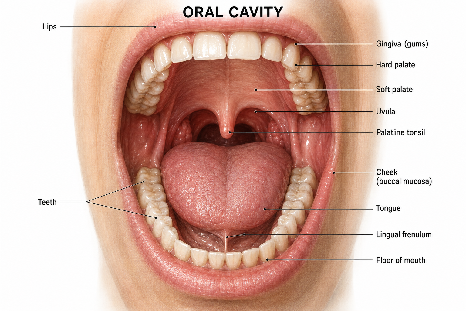
The oral cavity is the first part of the gastrointestinal tract. It is the region where food is received, mechanically broken down and mixed with saliva.
It is divided into the oral vestibule and oral cavity proper.
The oral cavity is bounded by the lips anteriorly, cheeks laterally, palate superiorly and tongue and floor of the mouth inferiorly.
3.2 Tongue
The tongue is a muscular organ that participates in mastication, taste, bolus formation and swallowing.
Its dorsal surface contains papillae. Filiform papillae mainly provide mechanical functions, while fungiform, foliate and circumvallate papillae are associated with taste sensation.
3.3 Teeth
Teeth mechanically break food into smaller particles during mastication. Permanent human dentition normally contains 32 teeth.
- Incisors – cutting
- Canines – tearing
- Premolars – crushing
- Molars – grinding
3.4 Functions of oral cavity
- Ingestion
- Mastication
- Mixing with saliva
- Bolus formation
- Initiation of carbohydrate digestion
- Taste
- Beginning of swallowing
3.5 Microscopic anatomy
Most of the oral cavity is lined by stratified squamous epithelium. In areas exposed to significant mechanical stress, such as the gingiva and hard palate, the epithelium is keratinized or parakeratinized. Other regions are generally lined by non-keratinized stratified squamous epithelium.
4. Pharynx
The pharynx is a muscular tube situated behind the nasal and oral cavities. It is shared by the respiratory and digestive systems. During swallowing, it provides a pathway through which the food bolus passes toward the esophagus.
Parts
- Nasopharynx
- Oropharynx
- Laryngopharynx
Functions
- Passage of food.
- Participation in swallowing.
- Protection of the lower respiratory tract during swallowing.
- Movement of food toward the esophagus.
Microscopic anatomy
The epithelium varies according to function. The nasopharynx is primarily lined by respiratory epithelium, whereas regions exposed to food are lined mainly by stratified squamous epithelium. The wall contains skeletal muscle involved in swallowing.
5. Esophagus
5.1 Gross anatomy
The esophagus is a muscular tube approximately 25 cm long in the adult. It extends from the pharynx to the stomach and passes through the neck and thorax before crossing the diaphragm.
It begins approximately at the level of the sixth cervical vertebra and joins the stomach at the gastroesophageal junction.

5.2 Functions
- Receives food from the pharynx.
- Transports food to the stomach.
- Uses peristaltic contractions.
- Participates in protection against gastroesophageal reflux.
5.3 Microscopic anatomy
The esophageal wall contains four basic layers.
Mucosa
The mucosa is lined by stratified squamous non-keratinized epithelium, which provides protection against mechanical abrasion.
Submucosa
The submucosa contains connective tissue, blood vessels, lymphatic vessels and esophageal glands that contribute to lubrication.
Muscularis externa
The upper third contains mainly skeletal muscle, the middle third contains both skeletal and smooth muscle, and the lower third contains predominantly smooth muscle.
Adventitia
Most of the esophagus is surrounded by adventitia. The short abdominal portion has a serosal covering.
6. Stomach
6.1 Gross anatomy
The stomach is a J-shaped muscular organ situated mainly in the upper left portion of the abdominal cavity. It lies between the esophagus and duodenum.
Parts of stomach
- Cardia – region surrounding the esophageal opening.
- Fundus – dome-shaped superior portion.
- Body – largest central region.
- Pyloric part – distal region leading to the duodenum.

6.2 Curvatures
- Lesser curvature
- Greater curvature
6.3 Sphincters
The lower esophageal region helps regulate entry of food into the stomach, while the pyloric sphincter regulates passage of gastric contents into the duodenum.
6.4 Functions
- Temporary storage of food.
- Mechanical mixing.
- Formation of chyme.
- Protein digestion.
- Secretion of hydrochloric acid.
- Secretion of intrinsic factor.
- Secretion of digestive enzymes.
- Regulation of gastric emptying.
6.5 Microscopic anatomy
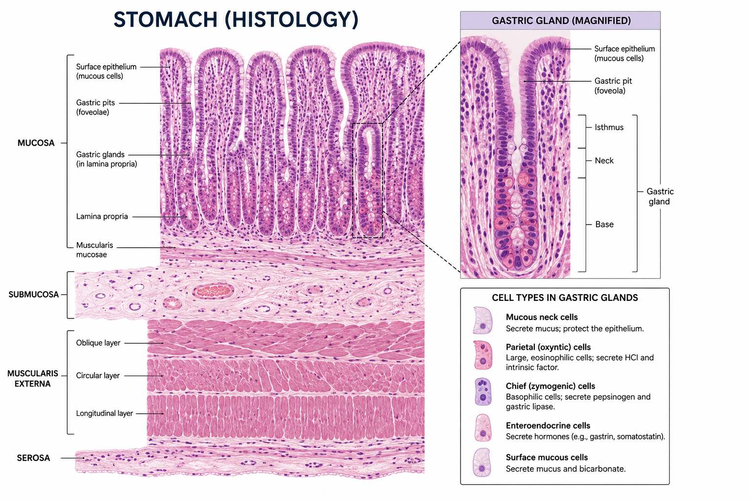
The gastric mucosa is lined by simple columnar epithelium. The surface epithelium forms gastric pits that open into gastric glands.
6.6 Gastric gland cells
| Cell | Major secretion | Importance |
|---|---|---|
| Mucous cells | Mucus | Protects gastric mucosa |
| Parietal cells | HCl and intrinsic factor | Acidification and vitamin B12 absorption |
| Chief cells | Pepsinogen | Protein digestion |
| G cells | Gastrin | Stimulates gastric secretion and motility |
| ECL cells | Histamine | Promotes acid secretion |
6.7 Mechanism of gastric acid secretion
Hydrogen ions are secreted by parietal cells through the H+/K+-ATPase located in the apical membrane. Chloride ions also enter the gastric lumen, resulting in the formation of hydrochloric acid.
Gastric acid secretion is stimulated by acetylcholine, gastrin and histamine. These mediators act together to increase activity of the proton pump.
6.8 Protein digestion in stomach
Chief cells secrete pepsinogen. In the acidic environment created by HCl, pepsinogen is converted to pepsin. Pepsin begins the digestion of proteins into smaller peptides.
7. Small Intestine
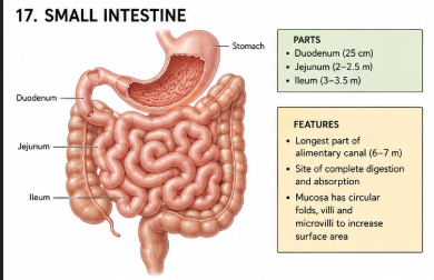
The small intestine is the longest part of the gastrointestinal tract and the principal site of digestion and nutrient absorption.
7.1 Parts
- Duodenum
- Jejunum
- Ileum
7.2 Duodenum
The duodenum is the first part of the small intestine. It receives acidic chyme from the stomach together with bile and pancreatic juice.
The duodenal mucosa contains Brunner's glands in the submucosa, which produce alkaline mucus that contributes to protection of the mucosa and neutralization of gastric acid.
7.3 Jejunum
The jejunum is the middle portion of the small intestine. It has prominent circular folds and long villi and is specialized for absorption of nutrients.
7.4 Ileum
The ileum is the terminal part of the small intestine and joins the cecum at the ileocecal junction. It contains prominent aggregations of lymphoid tissue known as Peyer's patches.
7.5 Microscopic anatomy
The small intestine contains several structural adaptations for absorption.
- Circular folds
- Villi
- Microvilli
- Intestinal crypts
- Enterocytes
- Goblet cells
- Paneth cells
- Enteroendocrine cells
7.6 Intestinal villus
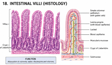
Each villus contains a network of blood capillaries and a central lymphatic vessel called a lacteal. Monosaccharides and amino acids are transported mainly into blood capillaries, whereas many products of long-chain lipid digestion are transported into lacteals as chylomicrons.
7.7 Functions
- Neutralization of gastric chyme.
- Digestion of carbohydrates.
- Digestion of proteins.
- Digestion of lipids.
- Absorption of nutrients.
- Absorption of water and electrolytes.
- Endocrine regulation.
- Immune defense.
8. Large Intestine
8.1 Parts
- Cecum
- Vermiform appendix
- Ascending colon
- Transverse colon
- Descending colon
- Sigmoid colon
- Rectum
- Anal canal

8.2 Characteristic features
- Taeniae coli
- Haustra
- Epiploic appendages
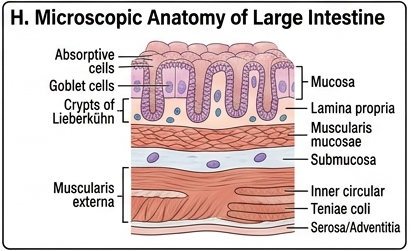
8.3 Microscopic anatomy
The mucosa of the large intestine is lined by simple columnar epithelium. It contains numerous straight intestinal crypts and abundant goblet cells.
A major distinguishing feature is the absence of intestinal villi.
8.4 Functions
- Absorption of water.
- Absorption of electrolytes.
- Formation of feces.
- Storage of feces.
- Support of intestinal microbiota.
- Absorption of some microbial metabolites.
9. Rectum and Anal Canal
Rectum
The rectum is the terminal expanded portion of the large intestine. It serves mainly as a temporary reservoir for feces before defecation.
Anal canal
The anal canal is the final part of the gastrointestinal tract. It contains internal and external anal sphincters that regulate defecation.
Defecation
Distension of the rectum stimulates stretch receptors. Signals travel through the nervous system and initiate the defecation reflex. Contraction of the rectal wall together with relaxation of the internal anal sphincter facilitates movement of feces toward the anal canal. Voluntary control of the external anal sphincter allows defecation to be consciously regulated.
10. Salivary Glands
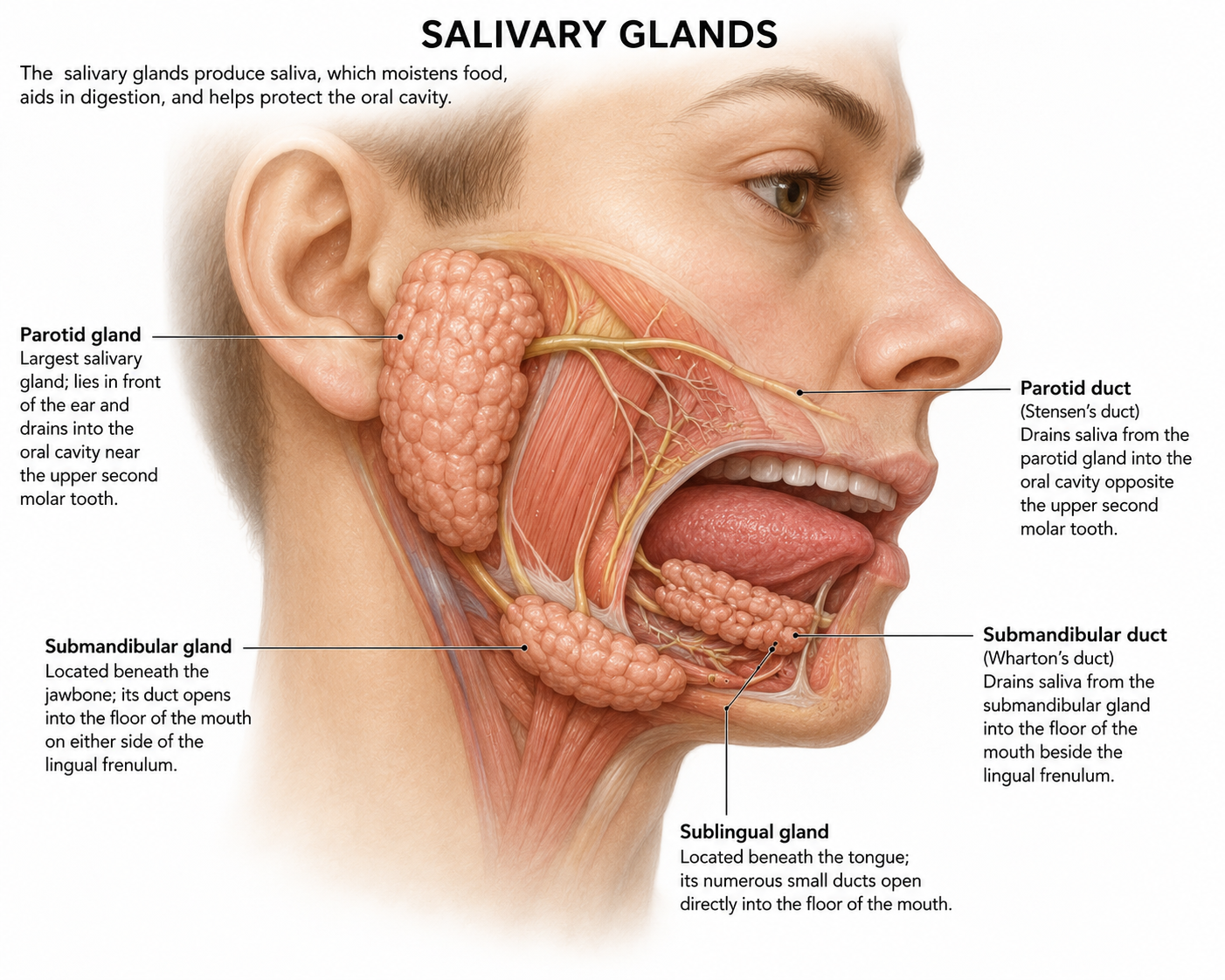
Salivary glands are exocrine glands that secrete saliva into the oral cavity. Saliva plays important roles in lubrication, digestion, taste, oral protection and maintenance of the oral environment.
10.1 Major salivary glands
| Gland | Location | Nature of secretion | Main features |
|---|---|---|---|
| Parotid | Below and anterior to ear | Predominantly serous | Rich in amylase |
| Submandibular | Below mandible | Mixed, predominantly serous | Major contributor to resting saliva |
| Sublingual | Floor of mouth | Mixed, predominantly mucous | Produces viscous mucus-rich secretion |
10.2 Microscopic structure
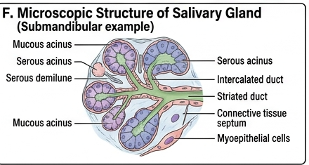
Salivary glands consist of secretory end pieces and a branching duct system. Secretory units may be serous, mucous or mixed.
Serous cells
Serous cells produce a watery protein-rich secretion containing enzymes such as salivary amylase.
Mucous cells
Mucous cells produce a viscous secretion rich in mucins. Mucins lubricate the oral cavity and food bolus.
Duct system
Secretions pass from secretory acini into intercalated ducts, then striated ducts and larger excretory ducts before reaching the oral cavity.
10.3 Functions of saliva
- Lubrication of food.
- Formation of food bolus.
- Initiation of starch digestion.
- Solubilization of substances for taste.
- Protection of oral mucosa.
- Antimicrobial activity.
- Buffering of oral acids.
- Maintenance of dental health.
10.4 Regulation of salivation
Salivary secretion is controlled mainly by the autonomic nervous system. Parasympathetic stimulation generally produces a copious watery secretion, whereas sympathetic stimulation can produce a smaller, more protein-rich secretion.
11. Pancreas
The pancreas is a mixed gland because it performs both exocrine and endocrine functions. Its exocrine component is essential for digestion, while its endocrine component regulates blood glucose and other metabolic processes.

11.1 Gross anatomy
The pancreas is an elongated organ located posterior to the stomach. It extends from the duodenum toward the spleen.
Parts
- Head
- Uncinate process
- Neck
- Body
- Tail
11.2 Exocrine pancreas
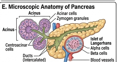
The exocrine pancreas is composed mainly of pancreatic acini and a duct system. Acinar cells synthesize and secrete digestive enzymes, many of which are produced initially as inactive precursors.
11.3 Pancreatic juice
Pancreatic juice contains digestive enzymes and bicarbonate. Bicarbonate is important for neutralizing acidic chyme entering the duodenum from the stomach.
| Enzyme/substance | Major function |
|---|---|
| Pancreatic amylase | Digestion of starch and glycogen |
| Pancreatic lipase | Digestion of triglycerides |
| Trypsinogen | Precursor of trypsin |
| Chymotrypsinogen | Precursor of chymotrypsin |
| Procarboxypeptidase | Precursor of carboxypeptidase |
| Nucleases | Digestion of nucleic acids |
| Bicarbonate | Neutralizes gastric acid |
11.4 Activation of pancreatic proteases
Pancreatic proteases are largely secreted as inactive zymogens. Enteropeptidase at the brush border of duodenal epithelial cells converts trypsinogen to trypsin. Trypsin then activates several other pancreatic protease zymogens.
11.5 Endocrine pancreas
The endocrine portion consists of the pancreatic islets or Islets of Langerhans. These are scattered among the exocrine acini.
| Cell | Hormone | Major function |
|---|---|---|
| Beta cells | Insulin | Promotes glucose uptake and lowers blood glucose |
| Alpha cells | Glucagon | Raises blood glucose |
| Delta cells | Somatostatin | Inhibits several endocrine and gastrointestinal secretions |
| PP cells | Pancreatic polypeptide | Participates in regulation of pancreatic and GI activity |
11.6 Functions of pancreas
- Digestion of carbohydrates.
- Digestion of proteins.
- Digestion of lipids.
- Digestion of nucleic acids.
- Neutralization of gastric acid.
- Regulation of blood glucose.
12. Liver
The liver is the largest internal gland of the human body and performs numerous metabolic, synthetic, storage, detoxification, excretory and immune functions.

12.1 Location
The liver lies mainly in the right upper quadrant of the abdomen immediately below the diaphragm, with a smaller portion extending toward the left side.
12.2 Surfaces
- Diaphragmatic surface
- Visceral surface
12.3 Anatomical lobes
The liver is anatomically divided into a large right lobe and a smaller left lobe. On the visceral surface, the caudate and quadrate lobes are also recognized.
12.4 Porta hepatis
The porta hepatis is a transverse fissure on the visceral surface through which major vessels, ducts and nerves enter or leave the liver.
Important structures associated with the porta hepatis include:
- Hepatic portal vein
- Hepatic artery
- Hepatic ducts
- Lymphatic vessels
- Nerves
12.5 Blood supply
The liver has a dual blood supply.
Hepatic portal vein
The portal vein carries nutrient-rich blood from the gastrointestinal tract and spleen to the liver.
Hepatic artery
The hepatic artery supplies oxygen-rich arterial blood to the liver tissue.
12.6 Venous drainage
Blood from hepatic sinusoids drains into central veins, then collecting veins and finally the hepatic veins, which drain into the inferior vena cava.
12.7 Microscopic anatomy of liver
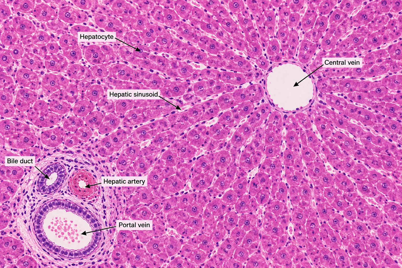
The basic microscopic organization of the liver can be described using the classical hepatic lobule, portal lobule and hepatic acinus.
12.8 Classical hepatic lobule
The classical hepatic lobule is approximately hexagonal and is organized around a central vein. Hepatocytes are arranged in plates or cords radiating from the central vein.
12.9 Portal triad
A portal triad is located within a portal area at the periphery of a classical lobule. It consists of branches of:
- Portal vein
- Hepatic artery
- Bile duct
12.10 Hepatocytes
Hepatocytes are the principal functional cells of the liver. They are involved in metabolism, synthesis, detoxification, bile formation, storage and processing of various substances.
12.11 Hepatic sinusoids
Hepatic sinusoids are specialized vascular channels located between plates of hepatocytes. Blood from branches of the hepatic artery and portal vein mixes in the sinusoids and flows toward the central vein.
12.12 Kupffer cells
Kupffer cells are resident macrophages associated with hepatic sinusoids. They remove cellular debris, aged cells and certain microorganisms and contribute to immune surveillance.
12.13 Space of Disse
The space of Disse is a narrow perisinusoidal space between sinusoidal endothelial cells and hepatocytes. It plays an important role in exchange of substances between blood and hepatocytes.
12.14 Bile canaliculi
Bile canaliculi are tiny channels formed between adjacent hepatocytes. They collect bile produced by hepatocytes and carry it toward bile ductules located in portal areas.
12.15 Direction of blood flow and bile flow
Blood flow
Portal venule and hepatic arteriole → sinusoids → central vein → collecting veins → hepatic veins
Bile flow
Hepatocytes → bile canaliculi → canals of Hering/bile ductules → interlobular bile ducts → hepatic ducts
12.16 Major functions of liver
- Carbohydrate metabolism: Storage of glucose as glycogen and regulation of blood glucose.
- Lipid metabolism: Oxidation of fatty acids, synthesis of cholesterol, lipoproteins and other lipids.
- Protein metabolism: Amino acid metabolism and synthesis of many plasma proteins.
- Bile production: Synthesis and secretion of bile.
- Detoxification: Biotransformation of drugs and various foreign substances.
- Storage: Storage of glycogen and several vitamins and minerals.
- Bilirubin metabolism: Processing and excretion of bilirubin through bile.
- Immune function: Kupffer cells contribute to removal of particles and microorganisms from portal blood.
13. Gall Bladder
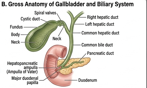
The gall bladder is a pear-shaped muscular sac situated on the visceral surface of the liver. It stores and concentrates bile produced by the liver.
13.1 Parts
- Fundus
- Body
- Neck
13.2 Biliary pathway
13.3 Microscopic anatomy
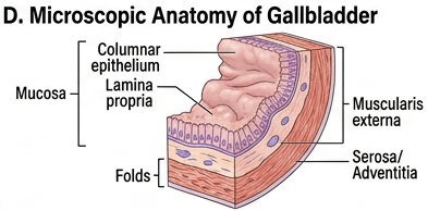
The wall of the gall bladder contains a mucosal lining, muscular layer and an outer connective tissue/serosal covering depending on the surface.
Mucosa
The mucosa is lined by simple columnar epithelial cells with microvilli. The mucosa forms numerous folds.
Muscular layer
The wall contains irregular bundles of smooth muscle that contract to expel bile.
Outer covering
The surface facing the peritoneal cavity is covered by serosa, while the surface attached to the liver has connective tissue (adventitial) covering.
13.4 Functions
- Storage of bile.
- Concentration of bile.
- Controlled release of bile into duodenum.
- Facilitation of fat digestion.
- Facilitation of absorption of dietary lipids and fat-soluble vitamins.
13.5 Regulation of gall bladder contraction
When fatty chyme enters the duodenum, enteroendocrine cells release cholecystokinin (CCK). CCK stimulates contraction of the gall bladder and promotes delivery of bile into the duodenum.
14. Mechanism of Digestion
Digestion is the process by which complex food substances are mechanically and chemically broken down into smaller molecules that can be absorbed.
Major stages
14.1 Digestion of carbohydrates
In mouth
Salivary amylase begins the digestion of starch and glycogen. It breaks internal alpha-1,4 glycosidic bonds and produces smaller carbohydrates such as maltose and dextrins.
In stomach
As food mixes with acidic gastric juice, salivary amylase becomes progressively inactive. Therefore, little carbohydrate digestion occurs in the stomach.
In small intestine
Pancreatic amylase continues carbohydrate digestion in the duodenum and jejunum.
Brush-border enzymes complete digestion.
| Enzyme | Substrate | Product |
|---|---|---|
| Maltase | Maltose | Glucose |
| Sucrase | Sucrose | Glucose + fructose |
| Lactase | Lactose | Glucose + galactose |
| Isomaltase | Isomaltose | Glucose |
14.2 Digestion of proteins
Stomach
Protein digestion begins in the stomach. Hydrochloric acid denatures proteins and activates pepsinogen to pepsin. Pepsin breaks proteins into smaller peptides.
Small intestine
Pancreatic proteases continue digestion. Trypsin, chymotrypsin, elastase and carboxypeptidases participate in breakdown of proteins and peptides.
Brush-border digestion
Peptidases associated with intestinal epithelial cells further break peptides into amino acids and small peptides.
14.3 Digestion of lipids
Most lipid digestion takes place in the small intestine. Because lipids are poorly soluble in water, bile salts are important for emulsification and formation of micelles.
Emulsification
Bile salts break large fat globules into smaller droplets, increasing the surface area available to pancreatic lipase.
Enzymatic digestion
Pancreatic lipase hydrolyses triglycerides mainly into monoglycerides and free fatty acids.
Micelle formation
Bile salts surround lipid digestion products and form micelles, which facilitate delivery of lipids to the intestinal epithelial surface.
Absorption
Long-chain fatty acids and monoglycerides enter enterocytes, where they are reassembled into triglycerides and packaged with other lipids and proteins into chylomicrons.
Chylomicrons enter intestinal lacteals and are transported through the lymphatic system before reaching the bloodstream.
14.4 Digestion of nucleic acids
Pancreatic nucleases digest DNA and RNA into smaller nucleotide components. Brush-border enzymes further process these products into absorbable bases, sugars and phosphate-containing molecules.
14.5 Complete digestion table
| Nutrient | Beginning of digestion | Major enzymes/substances | Final products | Main site of absorption |
|---|---|---|---|---|
| Carbohydrate | Mouth | Salivary and pancreatic amylase; disaccharidases | Monosaccharides | Small intestine |
| Protein | Stomach | Pepsin; pancreatic proteases; peptidases | Amino acids and small peptides | Small intestine |
| Lipid | Small amounts in mouth/stomach | Bile salts; pancreatic lipase | Fatty acids and monoglycerides | Small intestine |
| Nucleic acids | Small intestine | Pancreatic nucleases and intestinal enzymes | Bases, sugars and phosphate-containing products | Small intestine |
15. Absorption of Nutrients
Absorption is the movement of digested nutrients from the gastrointestinal lumen into blood or lymph. The small intestine is the principal site of nutrient absorption.
15.1 Carbohydrate absorption
Glucose and galactose are transported into enterocytes mainly through sodium-dependent cotransport mechanisms. Fructose is absorbed primarily by facilitated diffusion. Monosaccharides then enter intestinal blood vessels and pass through the hepatic portal circulation to the liver.
15.2 Protein absorption
Amino acids and small peptides are absorbed by specific transport systems in intestinal epithelial cells and enter the portal circulation.
15.3 Lipid absorption
Long-chain lipid digestion products enter enterocytes, are reassembled into triglycerides and packaged into chylomicrons. Chylomicrons enter lacteals and are transported through lymph.
15.4 Water absorption
Large quantities of water are absorbed throughout the intestine. Water follows osmotic gradients generated by absorption of solutes.
15.5 Vitamin and mineral absorption
Different vitamins and minerals are absorbed at different intestinal sites using different transport mechanisms. Vitamin B12 absorption depends on intrinsic factor and occurs mainly in the terminal ileum.
| Substance | Main absorption site/route |
|---|---|
| Glucose | Small intestine → portal blood |
| Amino acids | Small intestine → portal blood |
| Long-chain lipids | Small intestine → lacteals → lymph |
| Vitamin B12 | Terminal ileum with intrinsic factor |
| Water | Throughout intestine, especially small intestine and colon |
16. Deglutition and Gastrointestinal Motility
16.1 Deglutition
Deglutition, or swallowing, is the coordinated movement of food from the mouth to the stomach.
Stages of swallowing
- Oral stage: Voluntary movement of the bolus toward the pharynx.
- Pharyngeal stage: The swallowing reflex moves the bolus through the pharynx.
- Esophageal stage: Peristaltic contractions transport the bolus through the esophagus.
16.2 Peristalsis
Peristalsis is a coordinated wave of contraction and relaxation of gastrointestinal smooth muscle that propels luminal contents forward.
16.3 Segmentation
Segmentation consists of rhythmic contractions of intestinal smooth muscle that mix intestinal contents and bring them into contact with the absorptive mucosa.
17. Gastrointestinal Hormones
Gastrointestinal functions are regulated by neural, hormonal and local mechanisms.
| Hormone | Main source | Major action |
|---|---|---|
| Gastrin | G cells of stomach | Stimulates gastric acid secretion and gastric motility |
| Secretin | Duodenum | Stimulates pancreatic bicarbonate secretion |
| Cholecystokinin (CCK) | Duodenum and jejunum | Stimulates pancreatic enzyme secretion and gall bladder contraction |
| GIP | Small intestine | Enhances glucose-dependent insulin secretion and modulates gastric activity |
| Motilin | Small intestine | Promotes migrating motor activity |
18. Enteric Nervous System
The enteric nervous system is an extensive network of neurons within the wall of the gastrointestinal tract. It can coordinate many digestive activities independently, although it is strongly influenced by the autonomic nervous system.
Major plexuses
Submucosal plexus
The submucosal or Meissner's plexus is located in the submucosa and is particularly involved in regulation of secretion, blood flow and local mucosal activity.
Myenteric plexus
The myenteric or Auerbach's plexus lies between the circular and longitudinal muscle layers and is primarily involved in regulation of gastrointestinal motility.
19. Complete 10-Mark Long Questions
Introduction
The gastrointestinal system is a continuous muscular tube extending from the oral cavity to the anus, together with associated digestive glands and organs. It performs ingestion, digestion, absorption and elimination and also contributes to immune and endocrine functions.
Parts
The alimentary canal consists of the oral cavity, pharynx, esophagus, stomach, small intestine, large intestine, rectum and anal canal. Accessory organs include salivary glands, liver, gall bladder and pancreas.
General microscopic organization
The gastrointestinal wall generally contains mucosa, submucosa, muscularis externa and serosa or adventitia. The structure of the mucosa varies according to the specialized function of each region.
Gross and microscopic organization of individual parts
The oral cavity is specialized for ingestion and mastication. The esophagus is a muscular transport tube lined by protective stratified squamous epithelium. The stomach is a muscular reservoir containing gastric glands. The small intestine has villi and microvilli that maximize nutrient absorption. The large intestine lacks villi and contains numerous goblet cells for mucus secretion and fecal formation.
Functions
- Ingestion
- Mastication
- Swallowing
- Propulsion
- Secretion
- Mechanical digestion
- Chemical digestion
- Absorption
- Storage
- Defecation
Conclusion
The gastrointestinal system is structurally specialized along its length so that each region performs a particular role in the overall process of digestion and absorption.
Introduction
Digestion converts complex food substances into smaller molecules that can cross the intestinal epithelium. Carbohydrates are converted mainly into monosaccharides, proteins into amino acids and small peptides, and lipids into absorbable lipid products.
Carbohydrates
Digestion begins in the mouth through salivary amylase. Gastric acid later inhibits this enzyme. In the small intestine, pancreatic amylase continues digestion, while brush-border disaccharidases produce monosaccharides. Glucose and galactose are mainly absorbed by sodium-dependent transport, whereas fructose is absorbed mainly by facilitated diffusion. These monosaccharides enter portal blood.
Proteins
Protein digestion begins in the stomach. HCl denatures proteins and activates pepsinogen to pepsin. In the small intestine, pancreatic proteases including trypsin, chymotrypsin, elastase and carboxypeptidases continue digestion. Brush-border peptidases complete the process. Amino acids and small peptides are absorbed into portal blood.
Lipids
Most lipid digestion occurs in the small intestine. Bile salts emulsify dietary fat and pancreatic lipase hydrolyses triglycerides. The resulting products form micelles and enter enterocytes. Long-chain lipids are reassembled into triglycerides and packaged into chylomicrons, which enter intestinal lymphatics.
Conclusion
The coordinated action of gastrointestinal secretions, pancreatic enzymes, bile salts, intestinal enzymes and specialized absorptive structures allows efficient utilization of dietary nutrients.
Gross anatomy
The stomach is a J-shaped muscular organ located between the esophagus and duodenum. It consists of cardia, fundus, body and pyloric parts and has greater and lesser curvatures.
Microscopic anatomy
Its wall consists of mucosa, submucosa, muscularis externa and serosa. The mucosa contains gastric pits and glands lined by specialized cells. Mucous cells provide protection, parietal cells secrete HCl and intrinsic factor, chief cells secrete pepsinogen, and endocrine cells release regulatory hormones.
Functions
- Food storage
- Mixing
- Formation of chyme
- Protein digestion
- Acid secretion
- Intrinsic factor secretion
- Regulation of gastric emptying
Conclusion
The stomach combines mechanical mixing with chemical digestion and therefore acts both as a reservoir and a major digestive organ.
Gross anatomy
The small intestine consists of duodenum, jejunum and ileum. The duodenum receives bile and pancreatic juice, the jejunum is highly specialized for nutrient absorption, and the ileum continues absorption and contains prominent lymphoid tissue.
Microscopic anatomy
The mucosa contains circular folds, villi and microvilli. The epithelium consists mainly of absorptive enterocytes with goblet cells and specialized cells within intestinal crypts. The villi contain blood capillaries and lacteals.
Functions
- Completion of digestion
- Absorption of carbohydrates
- Absorption of proteins
- Absorption of lipids
- Absorption of vitamins and minerals
- Water and electrolyte absorption
- Endocrine regulation
- Immune protection
Gross anatomy
The large intestine consists of cecum, appendix, ascending colon, transverse colon, descending colon, sigmoid colon, rectum and anal canal. The colon has characteristic taeniae coli, haustra and epiploic appendages.
Microscopic anatomy
The mucosa contains simple columnar epithelium, numerous straight crypts and abundant goblet cells. Unlike the small intestine, villi are absent.
Functions
- Water absorption
- Electrolyte absorption
- Fecal formation
- Fecal storage
- Support of microbiota
- Absorption of microbial metabolites
Major glands
The three major paired salivary glands are parotid, submandibular and sublingual glands.
Structure
They are compound exocrine glands consisting of secretory acini and a duct system. Secretory units may be serous, mucous or mixed. The parotid is predominantly serous, the submandibular is mixed with predominantly serous secretion, and the sublingual is mixed with predominantly mucous secretion.
Functions
- Lubrication
- Bolus formation
- Starch digestion
- Taste
- Oral protection
- Antimicrobial action
- Buffering
Gross anatomy
The pancreas lies posterior to the stomach and consists of head, uncinate process, neck, body and tail. Its duct system opens into the duodenum.
Microscopic anatomy
The exocrine pancreas consists mainly of serous acini and ducts. The endocrine component consists of pancreatic islets scattered among the exocrine tissue.
Exocrine functions
Pancreatic juice contains amylase, lipase, protease zymogens, nucleases and bicarbonate. These substances are essential for digestion and neutralization of acidic chyme.
Endocrine functions
Islet cells secrete hormones including insulin, glucagon, somatostatin and pancreatic polypeptide.
Gross anatomy
The liver is located mainly in the right upper abdomen and has diaphragmatic and visceral surfaces. It has a large right lobe and smaller left lobe, with caudate and quadrate lobes recognized on the visceral surface.
Porta hepatis
The porta hepatis is the gateway through which major vessels, ducts, lymphatics and nerves enter or leave the liver.
Blood supply
The liver receives nutrient-rich blood from the portal vein and oxygenated blood from the hepatic artery. Blood passes through hepatic sinusoids toward central veins and eventually exits through hepatic veins.
Microscopic anatomy
The classical hepatic lobule is organized around a central vein. Hepatocyte plates radiate from the central vein and are separated by sinusoids. Portal areas at the periphery contain branches of the portal vein, hepatic artery and bile duct.
Other important structures
- Hepatocytes
- Sinusoids
- Kupffer cells
- Space of Disse
- Bile canaliculi
- Portal triads
Functions
- Carbohydrate metabolism
- Lipid metabolism
- Protein metabolism
- Bile production
- Detoxification
- Storage
- Bilirubin processing
- Plasma protein synthesis
- Immune function
Gross anatomy
The gall bladder is a pear-shaped organ located on the visceral surface of the liver. It consists of fundus, body and neck and connects with the biliary tree through the cystic duct.
Microscopic anatomy
The mucosa is lined by simple columnar epithelium and contains folds. The wall contains smooth muscle and an outer serosal or adventitial covering depending on its surface.
Functions
- Storage of bile
- Concentration of bile
- Controlled release of bile
- Assistance in lipid digestion
- Facilitation of absorption of dietary lipids
Regulation
CCK released from the small intestine in response to fatty intestinal contents stimulates gall bladder contraction and promotes bile delivery into the duodenum.
Introduction
Accessory digestive organs contribute secretions and mechanical functions required for efficient digestion.
Salivary glands
Produce saliva containing mucus, electrolytes and digestive enzymes, particularly salivary amylase.
Liver
Produces bile and performs major metabolic and detoxification functions.
Gall bladder
Stores and concentrates bile and releases it into the duodenum.
Pancreas
Produces pancreatic enzymes and bicarbonate for digestion and neutralization of gastric acid.
Conclusion
The accessory organs work together with the gastrointestinal tract to provide the secretions necessary for digestion, absorption and metabolic regulation.
Swallowing
Swallowing consists of oral, pharyngeal and esophageal phases. The oral phase is voluntary, while the pharyngeal and esophageal phases are largely reflexive.
Peristalsis
Peristalsis consists of coordinated contraction behind the food bolus and relaxation ahead of it. This produces forward movement through the gastrointestinal tract.
Segmentation
Segmentation produces mixing of intestinal contents and increases contact between nutrients and absorptive surfaces.
Importance
- Transport of food
- Mixing with digestive secretions
- Improved enzymatic digestion
- Improved contact with absorptive mucosa
| Region | Major digestive events | Major absorptive events |
|---|---|---|
| Mouth | Mastication and salivary digestion | Limited |
| Stomach | Protein digestion, mixing and acid secretion | Limited absorption |
| Duodenum | Bile and pancreatic digestion | Major nutrient absorption begins |
| Jejunum | Completion of digestion | Major nutrient absorption |
| Ileum | Continued digestion | Bile salts, vitamin B12 and other nutrients |
| Large intestine | Microbial activity | Water and electrolytes |
Overall digestion converts macromolecules into absorbable units, while specialized epithelial surfaces transport these nutrients into blood or lymph.
20. Separate 5-Mark Preparation
The stomach is a J-shaped muscular organ between the esophagus and duodenum. It consists of cardia, fundus, body and pyloric parts and has greater and lesser curvatures. It functions as a food reservoir, mixes food, produces chyme, begins protein digestion and secretes hydrochloric acid, pepsinogen and intrinsic factor. The pyloric region regulates gastric emptying.
The small intestinal wall consists of mucosa, submucosa, muscularis externa and serosa. The mucosa contains circular folds, villi and microvilli that increase absorptive surface area. Enterocytes perform absorption, goblet cells secrete mucus, Paneth cells contribute to antimicrobial defense and enteroendocrine cells secrete regulatory hormones. Villi contain blood capillaries and central lacteals.
The major salivary glands are parotid, submandibular and sublingual glands. The parotid is predominantly serous, the submandibular is mixed but predominantly serous, and the sublingual is mixed but predominantly mucous. Saliva lubricates food, helps form the bolus, begins starch digestion and protects the oral cavity.
The exocrine pancreas consists of acini and ducts. It secretes pancreatic juice containing amylase, lipase, protease zymogens, nucleases and bicarbonate. Amylase digests carbohydrates, lipase digests fats, proteases digest proteins and bicarbonate neutralizes acidic chyme entering the duodenum.
The classical hepatic lobule is approximately hexagonal and contains a central vein at its center. Hepatocytes are arranged in plates around the central vein, with hepatic sinusoids between the plates. Portal triads occur at the peripheral portal areas and contain branches of the portal vein, hepatic artery and bile duct. Kupffer cells occur within the sinusoidal system.
The gall bladder is a pear-shaped organ situated on the visceral surface of the liver. It consists of fundus, body and neck. It stores and concentrates bile and releases it into the duodenum following stimulation by CCK. Its mucosa is lined by simple columnar epithelium and contains folds.
Carbohydrate digestion begins in the mouth with salivary amylase. It is reduced in the stomach because of acidic pH and resumes in the small intestine through pancreatic amylase. Brush-border enzymes such as maltase, sucrase, lactase and isomaltase produce monosaccharides, which are absorbed mainly in the small intestine.
Protein digestion begins in the stomach through pepsin, which is activated in the presence of HCl. In the small intestine, trypsin, chymotrypsin, elastase and carboxypeptidases continue protein digestion. Brush-border peptidases produce absorbable amino acids and small peptides.
Most lipid digestion occurs in the small intestine. Bile salts emulsify fat and increase the surface available for pancreatic lipase. Lipase produces fatty acids and monoglycerides. These form micelles, enter enterocytes and are packaged into chylomicrons. Chylomicrons enter intestinal lymphatics.
The liver performs carbohydrate, lipid and protein metabolism, produces bile, synthesizes plasma proteins, stores glycogen and certain vitamins and minerals, participates in bilirubin metabolism, detoxifies many substances and contributes to immune defense through Kupffer cells.
- Absorption of water.
- Absorption of electrolytes.
- Formation of feces.
- Storage of feces.
- Support of intestinal microbiota.
- Absorption of some bacterial metabolites.
The GI tract generally consists of mucosa, submucosa, muscularis externa and serosa/adventitia. The mucosa is involved in secretion and absorption, the submucosa provides support and contains vessels and nerves, the muscularis externa produces mixing and propulsion, and the outer layer protects and anchors the organ.
21. Separate 2-Mark Preparation
| Question | Answer |
|---|---|
| Define gastrointestinal system. | It is the continuous muscular digestive tube from mouth to anus together with associated digestive organs. |
| Name the major parts of alimentary canal. | Oral cavity, pharynx, esophagus, stomach, small intestine, large intestine, rectum and anal canal. |
| Name the parts of small intestine. | Duodenum, jejunum and ileum. |
| Name the parts of large intestine. | Cecum, appendix, colon, rectum and anal canal. |
| Name the major salivary glands. | Parotid, submandibular and sublingual glands. |
| Name the parts of stomach. | Cardia, fundus, body and pyloric part. |
| Name the parts of pancreas. | Head, uncinate process, neck, body and tail. |
| Name the anatomical lobes of liver. | Right and left lobes; caudate and quadrate lobes are identified on the visceral surface. |
| What is porta hepatis? | It is the gateway on the visceral surface of liver through which major vessels, ducts, nerves and lymphatics enter or leave. |
| What is a hepatic lobule? | It is a structural microscopic unit of liver organized around a central vein and composed mainly of plates of hepatocytes. |
| What is a portal triad? | It consists of branches of the portal vein, hepatic artery and bile duct within a portal area. |
| Name two functions of liver. | Bile production and metabolism of nutrients. It also performs detoxification, storage, protein synthesis and immune functions. |
| What is the function of gall bladder? | It stores and concentrates bile and releases it into the duodenum. |
| Name two enzymes involved in carbohydrate digestion. | Salivary amylase and pancreatic amylase. |
| Name two enzymes involved in protein digestion. | Pepsin and trypsin. |
| What is the function of bile salts? | They emulsify dietary fats and facilitate lipid digestion and absorption. |
| What is peristalsis? | It is coordinated contraction and relaxation of GI smooth muscle that propels contents forward. |
| What is segmentation? | It is rhythmic intestinal contraction that mixes luminal contents and facilitates contact with the mucosa. |
| Name the four basic layers of GI tract. | Mucosa, submucosa, muscularis externa and serosa/adventitia. |
| Which part of GI tract has villi? | Small intestine. |
| Does large intestine contain villi? | No. Its mucosa contains numerous intestinal crypts and abundant goblet cells. |
| What is the function of lacteal? | It absorbs and transports dietary lipids, particularly chylomicrons, into the lymphatic system. |
| What is the function of parietal cells? | They secrete hydrochloric acid and intrinsic factor. |
| What is the function of chief cells? | They secrete pepsinogen. |
| What is the function of Kupffer cells? | They are hepatic macrophages involved in removal of particles, cellular debris and microorganisms. |
| What is CCK? | Cholecystokinin is a gastrointestinal hormone that stimulates gall bladder contraction and pancreatic enzyme secretion. |
| What is secretin? | Secretin is a duodenal hormone that promotes pancreatic bicarbonate secretion. |
22. Important Comparison Tables
Small Intestine vs Large Intestine
| Feature | Small intestine | Large intestine |
|---|---|---|
| Main function | Digestion and nutrient absorption | Water/electrolyte absorption and fecal formation |
| Villi | Present | Absent |
| Goblet cells | Present | Abundant |
| Major length | Longer | Shorter |
| Special feature | Villi and microvilli | Taeniae coli and haustra |
Parotid vs Submandibular vs Sublingual
| Feature | Parotid | Submandibular | Sublingual |
|---|---|---|---|
| Secretion | Serous | Mixed, mainly serous | Mixed, mainly mucous |
| Location | Near ear | Below mandible | Floor of mouth |
| Major role | Enzymatic secretion | Mixed secretion | Lubrication |
Exocrine vs Endocrine Pancreas
| Feature | Exocrine pancreas | Endocrine pancreas |
|---|---|---|
| Structure | Acini and ducts | Islets of Langerhans |
| Product | Digestive enzymes and bicarbonate | Hormones |
| Route | Ducts to duodenum | Bloodstream |
| Major role | Digestion | Metabolic regulation |
23. High-Yield Examination Points
- Small intestine = major site of digestion and absorption.
- Stomach = storage, mixing and initiation of protein digestion.
- Large intestine = water/electrolyte absorption and fecal formation.
- Villi and microvilli greatly increase intestinal absorptive surface area.
- Parietal cells = HCl + intrinsic factor.
- Chief cells = pepsinogen.
- Pancreatic acini = digestive enzymes.
- Pancreatic ducts = bicarbonate-rich secretion.
- Liver has dual blood supply: portal vein + hepatic artery.
- Portal triad = portal vein branch + hepatic artery branch + bile duct.
- Kupffer cells = hepatic macrophages.
- Gall bladder stores and concentrates bile.
- CCK stimulates gall bladder contraction.
- Secretin stimulates pancreatic bicarbonate secretion.
- Long-chain lipids are transported mainly through lymph as chylomicrons.
24. Important Diagrams to Practise for Examination
- Complete gastrointestinal tract
- General four-layer organization of GI tract
- Oral cavity and pharynx
- Esophagus – gross and microscopic structure
- Stomach – gross anatomy
- Stomach – gastric glands
- Small intestine – gross anatomy
- Small intestinal villus
- Large intestine – gross anatomy
- Salivary glands
- Salivary gland histology
- Pancreas – gross anatomy
- Pancreatic acinus and islet
- Liver – gross anatomy
- Classical hepatic lobule
- Portal triad
- Blood flow through hepatic lobule
- Bile flow through liver
- Gall bladder – gross anatomy
- Gall bladder histology
- Mechanism of carbohydrate digestion
- Mechanism of protein digestion
- Mechanism of lipid digestion
Definition/Introduction → Parts → Gross anatomy → Microscopic anatomy → Functions → Relevant mechanism/physiology → Labelled diagram → Conclusion
25. Rapid Revision Table
| Structure | Most important point |
|---|---|
| Oral cavity | Ingestion, mastication and initiation of carbohydrate digestion |
| Pharynx | Passage during swallowing |
| Esophagus | Peristaltic transport |
| Stomach | Storage, mixing and protein digestion |
| Duodenum | Receives bile and pancreatic juice |
| Jejunum | Major nutrient absorption |
| Ileum | Bile salt and vitamin B12 absorption; Peyer's patches |
| Large intestine | Water absorption and fecal formation |
| Salivary glands | Saliva secretion |
| Pancreas | Digestive enzymes + bicarbonate |
| Liver | Bile, metabolism, synthesis, storage and detoxification |
| Gall bladder | Bile storage and concentration |