Musculoskeletal System
Human Anatomy & Physiology — MB 551
MSc Medical Microbiology — II Semester
1. Introduction
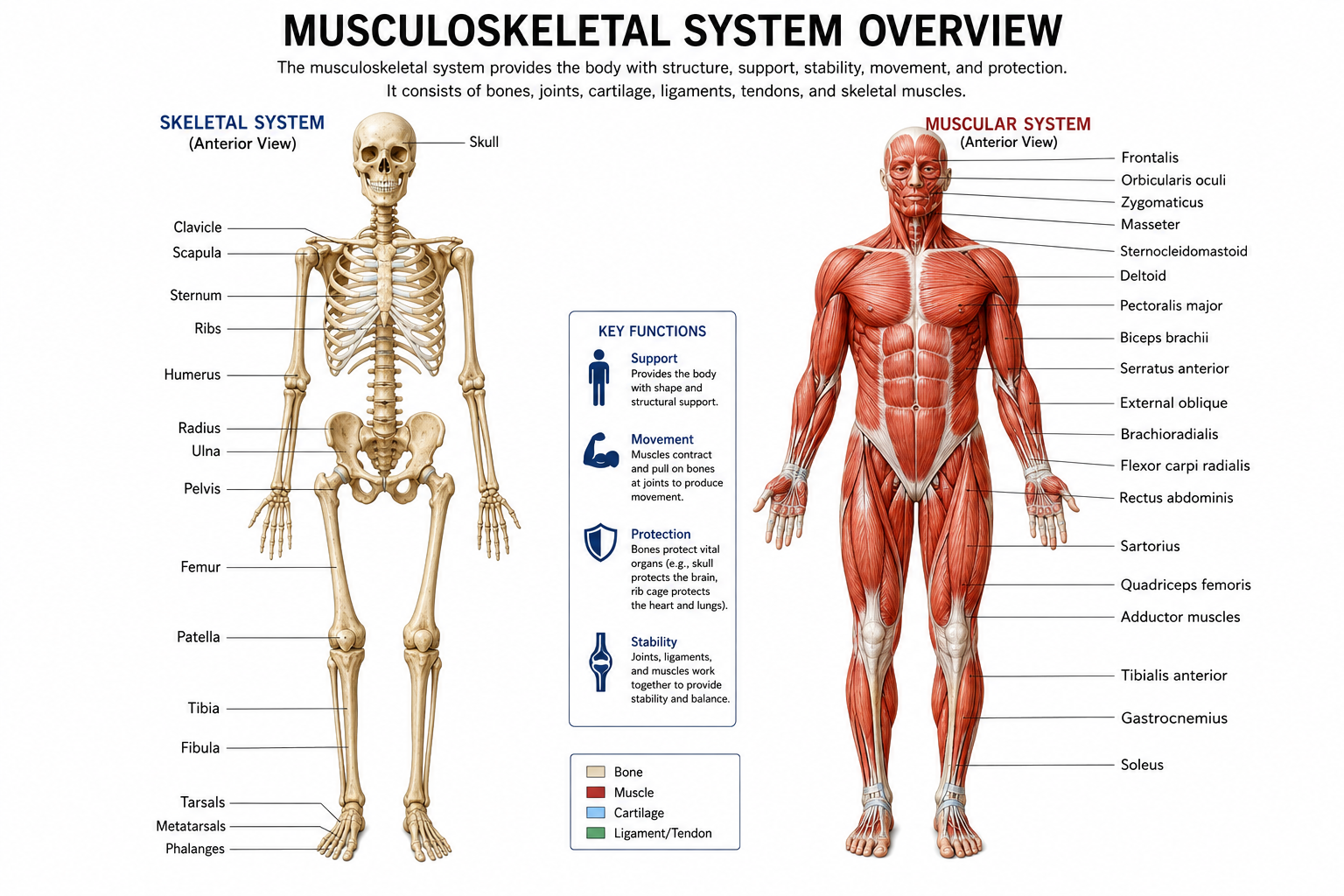
The musculoskeletal system is the body system responsible for support, stability, posture and movement. It consists mainly of bones, joints, skeletal muscles, cartilage, ligaments and tendons. These structures work together to provide a framework for the body and allow controlled movement.
Bones provide a rigid framework, muscles generate force, and joints permit movement between bones. The skeletal system also protects vital organs, stores minerals and contains bone marrow, which is involved in blood cell formation.
2. Skeleton
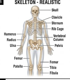
The human skeleton forms the structural framework of the body. In an adult, the skeleton consists of approximately 206 bones. It is divided into the axial skeleton and appendicular skeleton.
2.1 Axial Skeleton
The axial skeleton forms the central axis of the body. It includes the skull, vertebral column and thoracic cage.
- Skull
- Vertebral column
- Ribs
- Sternum
2.2 Appendicular Skeleton
The appendicular skeleton consists of the bones of the limbs and the pectoral and pelvic girdles that attach the limbs to the axial skeleton.
- Pectoral girdle
- Upper limbs
- Pelvic girdle
- Lower limbs
3. Classification of Bones
Bones can be classified according to their shape and structure. The major types are long, short, flat, irregular and sesamoid bones.
| Type of bone | Characteristics | Examples |
|---|---|---|
| Long bones | Longer than they are wide and mainly act as levers for movement. | Femur, humerus |
| Short bones | Small and approximately cuboidal in shape. | Carpals, tarsals |
| Flat bones | Thin and flattened bones that commonly protect internal organs. | Scapula, sternum, skull bones |
| Irregular bones | Bones with complex shapes that do not fit into the other categories. | Vertebrae |
| Sesamoid bones | Develop within tendons and help modify the mechanical action of muscles. | Patella |
4. Gross Anatomy of a Typical Long Bone
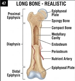
A typical long bone consists of a shaft called the diaphysis and expanded ends called epiphyses. Between the diaphysis and epiphysis is the metaphysis.
4.1 Diaphysis
The diaphysis is the elongated shaft of a long bone. It consists mainly of compact bone surrounding the medullary cavity.
4.2 Epiphysis
The epiphyses are the expanded ends of the bone. They contain mainly spongy bone with a thin outer layer of compact bone.
4.3 Metaphysis
The metaphysis is the region between the diaphysis and epiphysis. In growing individuals it contains the epiphyseal plate, which is responsible for longitudinal growth.
4.4 Periosteum
The periosteum is a connective tissue covering surrounding the external surface of bone except at articular surfaces. It contains blood vessels, nerves and cells involved in bone growth and repair.
4.5 Endosteum
The endosteum is a thin connective tissue membrane lining the internal surfaces of bone, including the medullary cavity and spaces within cancellous bone.
4.6 Medullary Cavity
The medullary cavity is the hollow central region of the diaphysis. In adults it mainly contains yellow bone marrow.
5. Microscopic Anatomy of Bone
Bone is a specialized connective tissue composed of cells embedded in a mineralized extracellular matrix. Microscopically, bone is classified mainly as compact bone and spongy bone.
5.1 Compact Bone
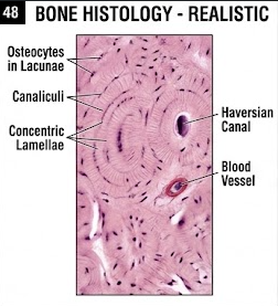
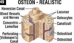
Compact bone is dense and forms the outer layer of most bones. Its structural unit is the osteon or Haversian system.
5.2 Osteon
An osteon consists of concentric layers of bone matrix called lamellae arranged around a central Haversian canal.
5.3 Spongy Bone
Spongy bone consists of thin plates or trabeculae arranged in a network. The spaces between trabeculae contain bone marrow.
6. Bone Cells and Their Functions
| Cell | Main function |
|---|---|
| Osteoblast | Produces new bone matrix and participates in bone formation. |
| Osteocyte | Maintains mature bone tissue and participates in regulation of bone metabolism. |
| Osteoclast | Resorbs bone tissue and participates in bone remodeling. |
| Osteoprogenitor cell | Acts as a precursor for osteoblasts. |
7. Bone Formation and Remodeling
Bone formation is called ossification or osteogenesis. It occurs mainly by intramembranous ossification and endochondral ossification.
7.1 Intramembranous Ossification
In intramembranous ossification, bone develops directly from mesenchymal tissue. This process contributes to the formation of many flat bones of the skull and parts of the clavicle.
7.2 Endochondral Ossification
Endochondral ossification involves replacement of a hyaline cartilage model by bone. It is the major process involved in formation of most long bones.
7.3 Bone Remodeling
Bone remodeling is a continuous process in which old bone is resorbed and new bone is formed. It helps maintain bone strength, repair microscopic damage and regulate mineral homeostasis.
8. Joints
A joint or articulation is a region where two or more bones meet. Joints provide varying degrees of movement and stability.
8.1 Fibrous Joints
Fibrous joints are connected by dense connective tissue and generally permit little or no movement.
8.2 Cartilaginous Joints
Cartilaginous joints are connected by cartilage and allow limited movement.
8.3 Synovial Joints
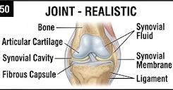
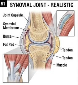
Synovial joints are freely movable joints characterized by a joint cavity, articular cartilage, synovial membrane and synovial fluid.
9. Structure of a Typical Synovial Joint
9.1 Articular Cartilage
Articular cartilage covers the articulating surfaces of bones. It reduces friction and helps distribute mechanical load.
9.2 Joint Capsule
The joint capsule encloses the joint cavity and consists of an outer fibrous layer and an inner synovial membrane.
9.3 Synovial Membrane
The synovial membrane lines the internal surface of the joint capsule and produces synovial fluid.
9.4 Synovial Fluid
Synovial fluid lubricates the joint and contributes to nutrition of the articular cartilage.
9.5 Ligaments
Ligaments connect bone to bone and help stabilize joints.
9.6 Tendons
Tendons connect muscles to bones and transmit the force generated by muscle contraction.
10. Muscles
Muscle tissue is specialized for contraction. The human body contains three major types of muscle tissue: skeletal muscle, cardiac muscle and smooth muscle.
| Type | Major features | Control |
|---|---|---|
| Skeletal muscle | Striated, multinucleated and attached mainly to bones. | Voluntary |
| Cardiac muscle | Striated muscle found in the heart with intercalated discs. | Involuntary |
| Smooth muscle | Non-striated muscle found in walls of hollow organs and blood vessels. | Involuntary |
11. Microscopic Anatomy of Skeletal Muscle
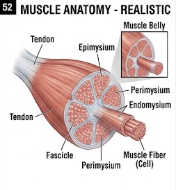
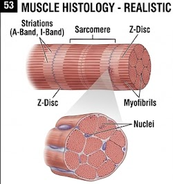
Skeletal muscle is composed of long cylindrical muscle fibers. Each muscle fiber contains numerous myofibrils, which consist of repeating functional units called sarcomeres.
11.1 Muscle Fiber
A skeletal muscle fiber is a long multinucleated cell surrounded by a plasma membrane called the sarcolemma.
11.2 Myofibrils
Myofibrils are contractile structures within muscle fibers. They contain actin and myosin filaments arranged in a regular pattern.
11.3 Sarcomere
The sarcomere is the basic functional contractile unit of skeletal and cardiac muscle. It extends between two Z discs.
12. Skeletal Muscle Contraction
Skeletal muscle contraction occurs through interaction between actin and myosin filaments. The process is explained by the sliding filament mechanism.
12.1 Neuromuscular Junction
The neuromuscular junction is the specialized synapse between a motor neuron and a skeletal muscle fiber. Acetylcholine acts as the neurotransmitter at this junction.

12.2 Sliding Filament Mechanism
- A motor nerve impulse reaches the neuromuscular junction.
- Acetylcholine is released from the motor nerve terminal.
- The muscle fiber membrane becomes depolarized.
- The action potential travels along the sarcolemma and transverse tubules.
- Calcium ions are released from the sarcoplasmic reticulum.
- Calcium binds to troponin and causes movement of tropomyosin.
- Myosin interacts with actin and produces filament sliding.
- Repeated cross-bridge cycling produces muscle contraction.
13. Clinical Correlations
13.1 Fracture
A fracture is a break or disruption in the continuity of a bone. It may occur because of trauma, excessive mechanical stress or weakened bone.
13.2 Osteoporosis
Osteoporosis is a condition characterized by reduced bone mass and deterioration of bone architecture, resulting in increased susceptibility to fractures.
13.3 Arthritis
Arthritis refers to disorders involving inflammation or degeneration of joints. Different forms can affect joint cartilage, synovium and surrounding structures.
13.4 Muscle Fatigue
Muscle fatigue refers to a decline in the ability of a muscle to generate force during sustained or repeated activity.
14. Important 10-Mark Long Questions
15. Important 5-Mark Questions
16. Important 2-Mark Questions
- Define osteon.
- What is an osteoblast?
- What is an osteoclast?
- Define synovial joint.
- What is synovial fluid?
- Define sarcomere.
- What is a neuromuscular junction?
- Name the three types of muscle tissue.
- What is the function of periosteum?
- What is bone remodeling?
17. High-Yield Revision
Bone
- Compact bone
- Spongy bone
- Osteon
- Bone cells
- Bone remodeling
Joints
- Fibrous
- Cartilaginous
- Synovial
- Joint capsule
- Synovial fluid
Muscle
- Skeletal muscle
- Muscle fiber
- Myofibril
- Sarcomere
- Actin and myosin
Contraction
- Neuromuscular junction
- Acetylcholine
- Calcium
- Troponin
- Sliding filament mechanism
18. Last-Minute Revision
- Osteoblast → bone formation
- Osteoclast → bone resorption
- Osteocyte → maintenance of mature bone
- Osteon → structural unit of compact bone
- Sarcomere → functional contractile unit of skeletal muscle
- Actin + myosin → responsible for filament interaction during contraction
- Synovial fluid → lubrication and nourishment of articular cartilage
- Tendon → connects muscle to bone
- Ligament → connects bone to bone
Recommended Medical Figures for Examination
The following figures are particularly useful for labelled diagrams and long-answer preparation.
Musculoskeletal system
Human skeleton
Long bone
Bone histology
Osteon
Joint anatomy
Synovial joint
Skeletal muscle
Muscle histology
Neuromuscular junction