Exam Alert:
The Endocrine System is a short syllabus unit in terms of teaching hours,
but it contains many highly testable concepts. For MSc-level preparation,
do not memorize only the names of hormones. Understand the
source → hormone → target → function → regulation →
hypo/hypersecretion disorder sequence.
MB 551
Human Anatomy & Physiology
3 hrs
Syllabus allocation
10-Mark
Long-question focus
High Yield
Hormones + disorders
Chapter Map
Table of Contents
Use the sections below for systematic exam preparation.
01
Definition and General Features of the Endocrine System
The endocrine system is a system of specialized cells,
tissues and glands that produce and release chemical messengers called
hormones into the blood or surrounding extracellular
fluid. These hormones travel to specific target cells or organs and
regulate physiological processes such as growth, metabolism,
reproduction, water and electrolyte balance, stress responses,
development and maintenance of internal homeostasis.
Unlike exocrine glands, endocrine glands generally do not possess ducts
through which their secretions are transported. They are therefore often
called ductless glands. Their secretory cells are usually
closely associated with a rich network of blood capillaries, allowing
hormones to enter the circulation efficiently.
Endocrine regulation is closely integrated with the nervous system.
The hypothalamus provides an important anatomical and functional link
between the nervous and endocrine systems. Through hypothalamic hormones
and neural signals, it regulates the pituitary gland, which in turn
controls several peripheral endocrine glands.
Important characteristics of endocrine glands
- They are generally ductless glands.
- They release hormones into blood or interstitial fluid.
- Hormones act on specific target cells possessing appropriate receptors.
- Many endocrine processes are controlled by negative feedback.
- Endocrine activity is important for maintaining homeostasis.
- Several endocrine organs also perform non-endocrine functions.
- The endocrine system interacts closely with the nervous and immune systems.
Endocrine gland versus exocrine gland
| Feature |
Endocrine gland |
Exocrine gland |
| Duct |
Usually absent |
Usually present |
| Secretion |
Hormones |
Enzymes, mucus, sweat, digestive secretions etc. |
| Route |
Blood/interstitial fluid |
Through ducts |
| Target |
Specific cells or organs with receptors |
Usually local surface or lumen |
| Examples |
Thyroid, pituitary, adrenal |
Salivary, sweat and exocrine pancreatic glands |
02
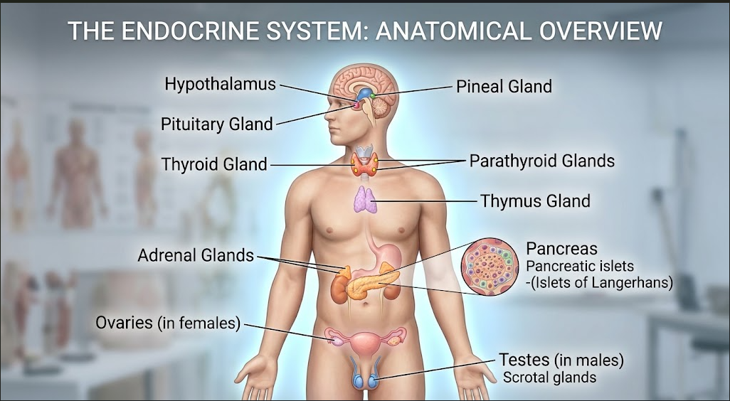
Major Endocrine Organs and Their Hormones
The endocrine system consists of several classical endocrine glands as
well as organs and tissues that possess endocrine functions. The major
glands important for MB 551 include the hypothalamus, pituitary, pineal,
thyroid, parathyroids, thymus, adrenal glands, endocrine pancreas,
testes and ovaries.
| Endocrine organ |
Major hormones |
Main physiological roles |
| Hypothalamus |
TRH, CRH, GnRH, GHRH, somatostatin, dopamine; synthesizes ADH and oxytocin |
Controls pituitary secretion, water balance, reproduction, growth and homeostasis |
| Pituitary |
GH, TSH, ACTH, FSH, LH, PRL, ADH, oxytocin |
Growth, thyroid function, adrenal function, reproduction, lactation and water balance |
| Pineal |
Melatonin |
Circadian and sleep-wake regulation |
| Thyroid |
T3, T4, calcitonin |
Metabolism, growth, development and calcium regulation |
| Parathyroid |
PTH |
Raises blood calcium and regulates phosphate balance |
| Thymus |
Thymic peptides including thymosin-related factors |
T-cell maturation and immune development |
| Adrenal cortex |
Aldosterone, cortisol, adrenal androgens |
Electrolyte balance, metabolism, stress response and androgenic effects |
| Adrenal medulla |
Epinephrine, norepinephrine |
Rapid sympathetic stress response |
| Pancreatic islets |
Insulin, glucagon, somatostatin, pancreatic polypeptide |
Glucose and energy homeostasis |
| Testes |
Testosterone, inhibin B, AMH during fetal life |
Male differentiation, spermatogenesis and reproductive function |
| Ovaries |
Estrogens, progesterone, inhibin, AMH |
Female reproductive cycle, pregnancy, secondary sex characteristics |
The hypothalamus is a small but functionally very important region of
the diencephalon. It lies below the thalamus and forms the floor and
part of the walls of the third ventricle. Although it is primarily a
neural structure, it has a major endocrine role because it regulates
the pituitary gland and synthesizes two posterior pituitary hormones:
antidiuretic hormone (ADH) and
oxytocin.
The hypothalamus acts as an important control centre for homeostasis.
It receives information related to body temperature, osmolarity,
energy status, stress and reproductive state and coordinates endocrine,
autonomic and behavioural responses.
Hypothalamic hormones
| Hormone |
Major action |
Target |
| TRH |
Stimulates TSH secretion and can also stimulate prolactin secretion |
Anterior pituitary |
| CRH |
Stimulates ACTH secretion |
Anterior pituitary |
| GnRH |
Stimulates secretion of FSH and LH |
Anterior pituitary |
| GHRH |
Stimulates GH secretion |
Anterior pituitary |
| Somatostatin |
Inhibits GH and TSH secretion; also has inhibitory effects in the gastrointestinal system |
Pituitary and other tissues |
| Dopamine |
Inhibits prolactin secretion |
Lactotrophs of anterior pituitary |
| ADH |
Promotes renal water reabsorption |
Kidney collecting ducts |
| Oxytocin |
Stimulates uterine contraction and milk ejection |
Uterus and mammary glands |
Important:
ADH and oxytocin are synthesized mainly in hypothalamic neurons,
transported down their axons and stored/released from the posterior
pituitary. Therefore, the posterior pituitary mainly serves as a
storage and release site rather than the site of hormone synthesis.
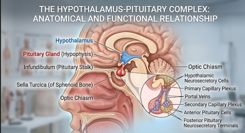
The pituitary gland, or hypophysis, is a small endocrine gland situated
at the base of the brain within the sella turcica of the
sphenoid bone. It is connected to the hypothalamus by the pituitary stalk.
Because several pituitary hormones regulate other endocrine glands,
the pituitary is often described as a major coordinating endocrine
gland. However, its activity itself is strongly controlled by the
hypothalamus and by feedback from peripheral hormones.
Anatomically and functionally, the pituitary is divided into an
anterior pituitary (adenohypophysis) and a
posterior pituitary (neurohypophysis). The anterior
pituitary is glandular tissue, whereas the posterior pituitary is
neural tissue associated with hypothalamic neurons.
Anterior pituitary hormones
| Hormone |
Major target |
Main functions |
| Growth hormone (GH) |
Liver, bone, cartilage, muscle and other tissues |
Promotes growth, protein synthesis and metabolic effects; many growth-promoting effects are mediated through IGF-1. |
| Thyroid-stimulating hormone (TSH) |
Thyroid gland |
Stimulates thyroid hormone synthesis and release and supports thyroid growth. |
| Adrenocorticotropic hormone (ACTH) |
Adrenal cortex |
Stimulates mainly cortisol synthesis and secretion. |
| Follicle-stimulating hormone (FSH) |
Ovaries/testes |
Promotes ovarian follicular development and supports spermatogenesis through Sertoli cells. |
| Luteinizing hormone (LH) |
Ovaries/testes |
Triggers ovulation and corpus luteum function in females and stimulates testosterone production by Leydig cells in males. |
| Prolactin (PRL) |
Mammary glands |
Promotes mammary gland development and milk production. |
Posterior pituitary hormones
| Hormone |
Main function |
| ADH / Vasopressin |
Increases water reabsorption in renal collecting ducts and contributes to maintenance of plasma osmolality and blood volume. At higher concentrations it also causes vasoconstriction. |
| Oxytocin |
Stimulates uterine contractions during labour and milk ejection during breastfeeding. |
Disorders of pituitary hormone secretion
Growth hormone deficiency
Deficiency of GH during childhood can result in reduced linear growth
and proportionate short stature. The severity depends on the cause,
timing and associated hormonal abnormalities. In adults, GH deficiency
may affect body composition, muscle mass, bone density and quality of
life.
Growth hormone excess
Excess GH before epiphyseal closure causes
gigantism, characterized by excessive linear growth.
Excess GH after epiphyseal closure produces
acromegaly, in which there is enlargement of the
hands, feet, jaw and facial bones together with soft-tissue changes.
Cushing disease
Cushing disease specifically refers to a pituitary ACTH-producing
adenoma that causes excess cortisol production by the adrenal cortex.
It should be distinguished from the broader term
Cushing syndrome, which includes all causes of
pathological cortisol excess.
Hyperprolactinemia
Excess prolactin can interfere with the reproductive axis. Depending
on sex and clinical context, it may produce inappropriate milk
secretion, menstrual disturbance, infertility and reduced gonadal
function. Dopamine normally suppresses prolactin secretion, so
disruption of dopaminergic inhibition can contribute to elevated
prolactin.
Central diabetes insipidus
Deficiency of ADH secretion or impaired release from the
hypothalamic-pituitary system reduces renal water reabsorption.
Patients develop excessive production of dilute urine
(polyuria) and compensatory excessive thirst
(polydipsia).
SIADH
Inappropriate excess ADH activity causes excessive water retention,
producing dilutional hyponatremia. Clinical manifestations may range
from nausea and headache to neurological symptoms when hyponatremia is
severe or develops rapidly.
The pineal gland is a small endocrine structure located near the roof of
the third ventricle in the epithalamic region of the brain. Its major
endocrine secretion is melatonin, which is synthesized
primarily by pinealocytes.
Melatonin
Melatonin secretion is strongly related to the light-dark cycle.
Darkness promotes melatonin secretion, whereas exposure to light
suppresses it. The retina communicates light information to the
suprachiasmatic nucleus of the hypothalamus, which influences sympathetic
pathways reaching the pineal gland.
| Hormone |
Source |
Function |
| Melatonin |
Pinealocytes |
Helps synchronize circadian rhythms and sleep-wake timing. |
Clinical significance
Disturbance of the light-dark cycle, shift work, jet lag and certain
neurological conditions can alter circadian rhythmicity. Pineal region
masses may also produce neurological manifestations because of their
anatomical location. Changes in melatonin signalling can influence
sleep and circadian timing.
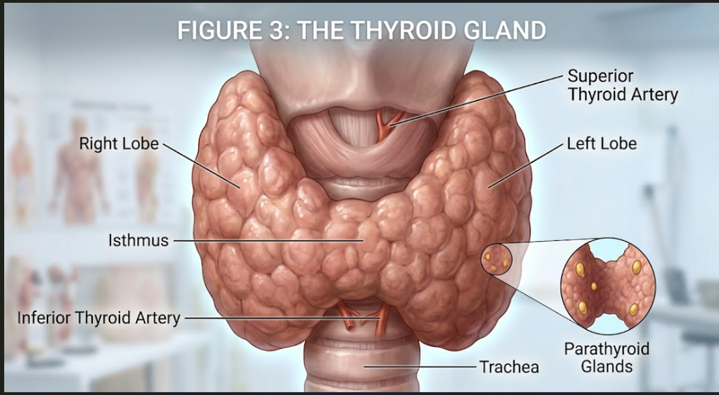
The thyroid gland is a highly vascular endocrine gland located in the
anterior neck, consisting of right and left lobes connected by an
isthmus. It lies anterior and lateral to the upper trachea and lower
laryngeal region.
Microscopically, the thyroid is composed of numerous spherical
thyroid follicles. Each follicle is lined mainly by
follicular cells and contains colloid rich in
thyroglobulin. Parafollicular or C cells occur between
follicles and produce calcitonin.
Hormones of the thyroid gland
| Hormone |
Cell of origin |
Major function |
| Thyroxine (T4) |
Follicular cells |
Major circulating thyroid hormone; serves as an important precursor for T3 and contributes to regulation of metabolism, growth and development. |
| Triiodothyronine (T3) |
Follicular cells; much formed by peripheral conversion of T4 |
More biologically active thyroid hormone; increases metabolic activity and supports normal growth and development. |
| Calcitonin |
Parafollicular C cells |
Helps reduce plasma calcium, particularly by inhibiting osteoclast-mediated bone resorption. |
Synthesis of thyroid hormones
Thyroid hormone synthesis is a complex process involving iodide uptake,
oxidation, organification and coupling reactions within the thyroid
follicle. Iodide is actively transported into follicular cells and is
then moved toward the follicular lumen. Thyroid peroxidase participates
in oxidation of iodide and its incorporation into tyrosine residues of
thyroglobulin.
-
Iodide trapping:
Follicular cells actively transport iodide from the blood.
-
Oxidation:
Iodide is converted to an activated form suitable for incorporation
into tyrosine residues.
-
Organification:
Iodinated tyrosine residues form MIT and DIT.
-
Coupling:
MIT and DIT residues combine to form thyroid hormones. DIT + DIT
produces T4, whereas MIT + DIT produces T3.
-
Storage:
Thyroid hormones remain stored within thyroglobulin in the colloid.
-
Release:
Thyroglobulin is taken back into follicular cells and processed to
release T3 and T4 into the circulation.
Functions of thyroid hormones
Thyroid hormones increase the metabolic activity of many tissues.
They influence oxygen consumption, heat production and utilization of
carbohydrates, fats and proteins. Their effects are particularly
important during growth and development, including normal development
of the central nervous system.
Thyroid hormones also increase the responsiveness of many tissues to
catecholamines by increasing the expression and functional effects of
adrenergic receptors. This contributes to cardiovascular manifestations
of thyroid hormone excess.
Regulation of thyroid secretion
Hypothalamus
→
TRH
→
Anterior pituitary
→
TSH
→
Thyroid
→
T3 + T4
Circulating T3 and T4 exert negative feedback on both the hypothalamus
and anterior pituitary. Therefore, when thyroid hormone levels increase,
TRH and TSH secretion generally decreases.
Hypothyroidism
Hypothyroidism is a state in which thyroid hormone
action is insufficient for the body's physiological requirements.
Reduced thyroid hormone action lowers metabolic activity and may produce
fatigue, cold intolerance, weight gain, constipation, dry skin and
slowed physiological processes.
In children, adequate thyroid hormone is particularly important for
normal growth and brain development. Severe untreated congenital
hypothyroidism can therefore cause serious developmental impairment.
Common causes include autoimmune thyroid disease such as
Hashimoto thyroiditis, iodine deficiency in susceptible
populations, thyroid gland destruction and disorders affecting
hypothalamic-pituitary stimulation.
Hyperthyroidism
Hyperthyroidism refers to excessive production and
secretion of thyroid hormones. Increased metabolic activity may result
in weight loss despite appetite, heat intolerance, sweating, tremor,
anxiety, tachycardia and increased bowel activity.
Graves disease is an important autoimmune cause of
hyperthyroidism. In this condition, antibodies stimulate the TSH receptor,
causing excessive thyroid hormone production. Some patients also develop
thyroid-associated ophthalmopathy.
| Condition |
Hormonal state |
Important features |
| Hypothyroidism |
Low thyroid hormone action |
Fatigue, cold intolerance, weight gain, constipation, dry skin, slowed metabolism |
| Hyperthyroidism |
Excess thyroid hormone action |
Heat intolerance, weight loss, tremor, sweating, tachycardia, increased metabolism |
| Goiter |
Thyroid enlargement; hormonal state may vary |
Neck swelling; may occur with hypo-, hyper- or normal thyroid function depending on cause |
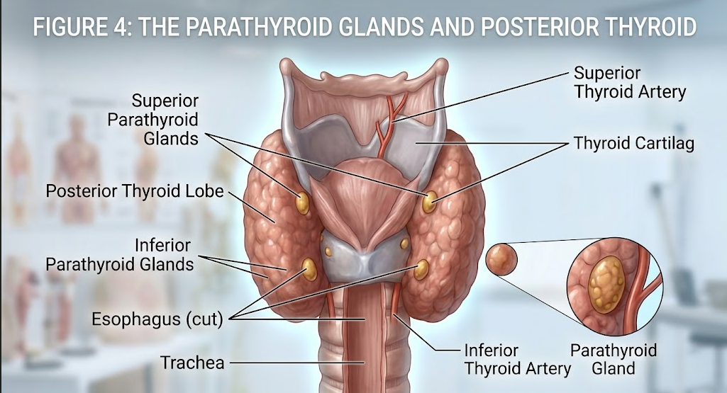
The parathyroid glands are small endocrine glands usually located on the
posterior aspect of the thyroid gland. Most individuals have four
glands, although the number and exact anatomical location may vary.
Parathyroid hormone
The major hormone of the parathyroid gland is
parathyroid hormone (PTH). Its principal physiological
function is to maintain extracellular calcium concentration. PTH is
released when blood calcium concentration falls.
| Site |
Effect of PTH |
| Bone |
Promotes bone resorption indirectly through osteoblast-mediated stimulation of osteoclast activity, increasing calcium release into blood. |
| Kidney |
Increases calcium reabsorption and decreases phosphate reabsorption, thereby increasing phosphate excretion. |
| Vitamin D metabolism |
Stimulates renal formation of active vitamin D, calcitriol. |
| Intestine |
Indirectly increases calcium absorption through increased calcitriol formation. |
Hypoparathyroidism
Hypoparathyroidism results in inadequate PTH activity and therefore
reduced serum calcium. Hypocalcemia increases neuromuscular excitability
and may produce tingling, muscle cramps, carpopedal spasm and
tetany. Severe hypocalcemia can cause seizures and
cardiac rhythm disturbances.
Hyperparathyroidism
Excess PTH causes increased calcium mobilization from bone, increased
renal calcium conservation and increased formation of calcitriol.
Persistent hyperparathyroidism can therefore produce
hypercalcemia.
Classical clinical manifestations of significant hypercalcemia include
renal stones, bone pain and skeletal weakening. Long-standing disease
can also affect renal function.
The thymus is a lymphoepithelial organ located in the anterior superior
mediastinum behind the sternum. It has both structural and endocrine-like
functions. It is particularly important during childhood, when it
provides the microenvironment required for maturation and selection of
T lymphocytes.
Thymic hormones and factors
Thymic epithelial cells produce several peptide factors, traditionally
described using terms such as thymosin and related
thymic hormones. These factors participate in T-cell development and
immune maturation.
| Endocrine function |
Importance |
| Thymic peptide factors |
Support maturation and differentiation of T lymphocytes. |
| Immune education |
Provides an environment for selection of developing T cells and establishment of appropriate immune tolerance. |
The thymus is relatively large and active during childhood and gradually
undergoes involution after puberty, with increasing replacement by
adipose tissue. However, thymic tissue can retain immune functions
throughout adult life.
Clinical correlation:
Disorders affecting thymic development can interfere with T-cell
maturation and cellular immunity. Thymic tumors such as thymoma are
clinically important, although they should not simply be classified as
classical hormone hypersecretion disorders.
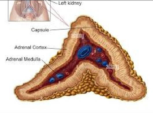
The adrenal glands are paired endocrine glands located superior to the
kidneys. Each gland consists of an outer adrenal cortex
and an inner adrenal medulla. These two regions differ
in embryological origin, histological organization and hormonal
function.
Adrenal cortex
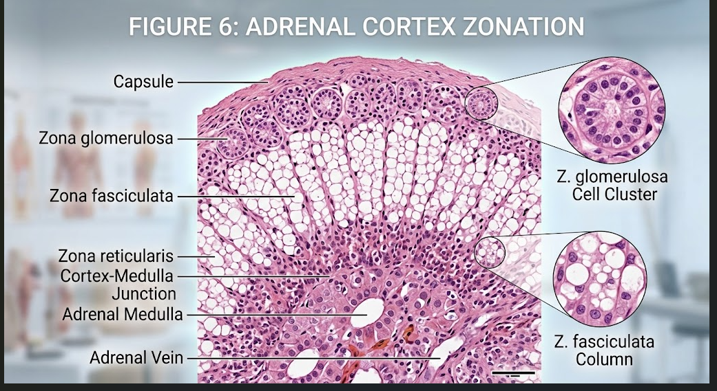
The adrenal cortex is divided into three concentric zones:
zona glomerulosa, zona fasciculata and zona reticularis.
A useful examination mnemonic is:
GFR = Salt, Sugar, Sex.
| Zone |
Major hormone |
Main function |
Main regulation |
| Zona glomerulosa |
Aldosterone |
Promotes sodium retention and potassium/hydrogen ion secretion; helps regulate extracellular fluid volume and blood pressure. |
Renin-angiotensin-aldosterone system and plasma potassium |
| Zona fasciculata |
Cortisol |
Supports metabolism and stress responses and has important anti-inflammatory and immunomodulatory actions. |
ACTH |
| Zona reticularis |
Adrenal androgens |
Contribute to androgenic activity, including development of some pubic and axillary hair and libido, particularly in females. |
ACTH contributes |
Aldosterone
Aldosterone is the major mineralocorticoid secreted by the zona
glomerulosa. It acts mainly on the distal nephron of the kidney,
increasing sodium reabsorption and increasing potassium and hydrogen
ion secretion. Because water follows retained sodium, aldosterone
contributes to maintenance of extracellular fluid volume and blood
pressure.
Cortisol
Cortisol is the major glucocorticoid produced by the zona fasciculata.
It has broad effects on metabolism and stress adaptation. Cortisol
increases hepatic gluconeogenesis, helps maintain blood glucose during
fasting and stress, influences protein and lipid metabolism and
suppresses excessive inflammatory and immune responses.
Cortisol secretion is regulated by the
hypothalamic-pituitary-adrenal axis. CRH from the
hypothalamus stimulates ACTH release from the anterior pituitary, and
ACTH stimulates cortisol secretion.
Adrenal androgens
The adrenal cortex produces weak androgens such as
dehydroepiandrosterone (DHEA) and related steroids. They contribute to
androgenic effects and serve as precursors for sex steroid synthesis in
peripheral tissues.
Adrenal medulla
The adrenal medulla contains chromaffin cells that are functionally
related to sympathetic postganglionic neurons. It secretes the
catecholamines epinephrine and norepinephrine.
| Hormone |
Major effects |
| Epinephrine |
Increases heart rate and contractility, promotes bronchodilation and mobilizes energy substrates during acute stress. |
| Norepinephrine |
Produces prominent vasoconstriction and contributes to increased vascular resistance and blood pressure. |
Adrenal hyposecretion disorders
Addison disease / Primary adrenal insufficiency
Primary adrenal insufficiency occurs when the adrenal cortex cannot
produce adequate steroid hormones, particularly cortisol and often
aldosterone. Reduced cortisol contributes to weakness, fatigue and
impaired stress tolerance. Aldosterone deficiency can cause sodium
loss, volume depletion and hyperkalemia.
In primary adrenal insufficiency, reduced cortisol feedback leads to
increased ACTH. Increased ACTH-related melanocortin activity may cause
characteristic hyperpigmentation.
Adrenal hypersecretion disorders
Cushing syndrome
Cushing syndrome is the clinical state caused by chronic excessive
glucocorticoid exposure. Causes include endogenous cortisol excess or
prolonged therapeutic glucocorticoid use. Features may include
central adiposity, muscle weakness, skin thinning, easy bruising,
hypertension and impaired glucose regulation.
Primary hyperaldosteronism
Excess aldosterone causes increased sodium retention and increased
potassium excretion. This may produce hypertension and hypokalemia.
Primary hyperaldosteronism is sometimes called
Conn syndrome.
Pheochromocytoma
Pheochromocytoma is a catecholamine-secreting tumor of chromaffin
tissue, usually arising in the adrenal medulla. Excess catecholamines
can produce episodic or persistent hypertension, headache, sweating,
palpitations and tremor.
10
Pancreas as an Endocrine Organ
The pancreas is a mixed gland because it has both exocrine and endocrine
components. The exocrine pancreas produces digestive enzymes, whereas
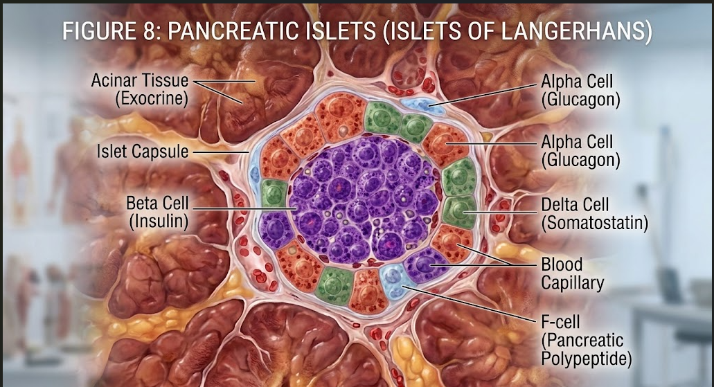
the endocrine component consists mainly of the
islets of Langerhans.
Pancreatic islets are scattered throughout the pancreatic tissue and
contain several endocrine cell types. These cells release hormones
directly into the blood and play a central role in glucose and energy
homeostasis.
Hormones of pancreatic islets
| Cell |
Hormone |
Main function |
| Beta cells |
Insulin |
Decreases blood glucose by promoting glucose uptake and storage and by inhibiting excessive hepatic glucose production. |
| Alpha cells |
Glucagon |
Raises blood glucose by stimulating hepatic glycogenolysis and gluconeogenesis. |
| Delta cells |
Somatostatin |
Inhibits secretion of insulin and glucagon and suppresses several gastrointestinal functions. |
| PP/Gamma cells |
Pancreatic polypeptide |
Influences pancreatic exocrine secretion, gastrointestinal activity and appetite-related processes. |
Insulin
Insulin is an anabolic hormone that is released mainly when blood
glucose concentration increases after a meal. It promotes glucose
uptake into insulin-sensitive tissues, particularly skeletal muscle
and adipose tissue, and promotes storage of energy as glycogen and
fat. It also supports protein synthesis.
Glucagon
Glucagon acts largely in opposition to insulin with respect to blood
glucose regulation. When blood glucose falls, glucagon stimulates the
liver to break down glycogen and synthesize new glucose. This helps
maintain plasma glucose during fasting.
Diabetes mellitus
Diabetes mellitus is a group of metabolic disorders characterized by
chronic hyperglycemia resulting from inadequate insulin secretion,
impaired insulin action or both.
| Feature |
Type 1 diabetes mellitus |
Type 2 diabetes mellitus |
| Major mechanism |
Usually autoimmune destruction of pancreatic beta cells |
Insulin resistance with progressive beta-cell dysfunction |
| Insulin secretion |
Markedly reduced or absent |
Initially may be preserved or increased, later becoming inadequate |
| Insulin resistance |
Not the primary mechanism |
Major component |
| Clinical consequence |
Hyperglycemia and tendency to ketosis without insulin treatment |
Chronic hyperglycemia with progressive metabolic and vascular complications |
Clinical point:
Persistent hyperglycemia can damage blood vessels and multiple organs,
including the kidneys, retina, nerves and cardiovascular system.
Therefore, endocrine dysfunction can produce important systemic
complications.
11
Testes as Endocrine Organs
The testes are male reproductive organs with both reproductive and
endocrine functions. Their endocrine activity is mainly carried out by
Leydig cells and Sertoli cells.
Leydig cells
Leydig cells are located in the interstitial tissue between seminiferous
tubules. Under stimulation by LH, they synthesize and secrete
testosterone.
Sertoli cells
Sertoli cells are located within the seminiferous epithelium and provide
structural and nutritional support to developing germ cells. They
produce inhibin B, which contributes to regulation of
FSH secretion, and produce anti-Müllerian hormone (AMH)
during fetal male development.
| Hormone |
Source |
Major functions |
| Testosterone |
Leydig cells |
Male differentiation, development of secondary sex characteristics, libido, anabolic effects and support of spermatogenesis. |
| Inhibin B |
Sertoli cells |
Provides negative feedback on FSH secretion and reflects Sertoli-cell/spermatogenic activity. |
| AMH |
Sertoli cells during fetal development |
Promotes regression of Müllerian ducts during male fetal differentiation. |
Hypogonadism
Testicular hypofunction may result in inadequate testosterone production,
impaired spermatogenesis or both. Depending on age of onset, it can
result in delayed or incomplete puberty, reduced secondary sexual
characteristics, reduced libido, infertility and changes in muscle and
bone mass.
Hypogonadism may be primary, due to a disorder of the
testes, or secondary, due to inadequate hypothalamic or
pituitary stimulation.
Excess androgen production
Excess androgen activity can result in virilization or inappropriate
sexual maturation, depending on the age and underlying cause.
Androgen-secreting tumors are an uncommon example.
12
Ovaries as Endocrine Organs
The ovaries are female reproductive organs with both gamete-producing
and endocrine functions. Important ovarian hormones include
estrogens, progesterone, inhibin and anti-Müllerian hormone.
Major ovarian hormones
| Hormone |
Main source |
Main functions |
| Estrogens |
Developing follicles and other ovarian tissues |
Female reproductive development, secondary sex characteristics, endometrial proliferation and maintenance of bone mass. |
| Progesterone |
Corpus luteum; placenta during pregnancy |
Transforms and maintains secretory endometrium, supports pregnancy and reduces uterine contractility. |
| Inhibin |
Granulosa cells |
Suppresses FSH secretion. |
| AMH |
Granulosa cells of developing follicles |
Regulates follicular recruitment and is clinically used as a marker related to ovarian reserve. |
Estrogen
Estrogens are important for development and maintenance of the female
reproductive system and secondary sex characteristics. They stimulate
proliferation of the endometrium during the proliferative phase of the
menstrual cycle and contribute to maintenance of bone mass.
Progesterone
Progesterone is particularly important after ovulation, when the corpus
luteum secretes it. It converts the estrogen-primed endometrium into a
secretory state suitable for implantation and helps maintain conditions
required for early pregnancy.
Ovarian endocrine dysfunction
Reduced ovarian hormone production can cause menstrual irregularity,
infertility, hot flashes and other symptoms associated with ovarian
insufficiency or menopause. Prolonged estrogen deficiency is also
associated with increased bone loss.
Excess estrogen or abnormal ovarian steroid production can contribute
to abnormal uterine bleeding and other reproductive disturbances,
depending on the underlying cause. Some ovarian tumors can also produce
steroid hormones.
Important distinction:
Polycystic ovary syndrome (PCOS) is a complex endocrine-metabolic and
reproductive disorder involving altered ovarian and adrenal androgen
activity, insulin resistance in many patients and abnormal
hypothalamic-pituitary-ovarian signalling. It should not be reduced to
simple “excess secretion of one ovarian hormone.”
13
Placenta and Other Endocrine Tissues
In addition to classical endocrine glands, several organs contain cells
capable of endocrine secretion. These tissues demonstrate that hormonal
regulation is distributed throughout the body.
Placenta
During pregnancy, the placenta acts as an important temporary endocrine
organ. It produces hormones that support implantation, maintenance of
pregnancy and preparation of the maternal body for lactation.
| Hormone |
Main function |
| hCG |
Maintains corpus luteum function during early pregnancy and supports progesterone production. |
| Estrogens |
Support uterine growth, reproductive tissue changes and preparation for lactation. |
| Progesterone |
Supports the endometrium and helps maintain pregnancy. |
| Human placental lactogen |
Alters maternal metabolism and contributes to preparation of the mammary glands for lactation. |
Other endocrine tissues
| Organ/tissue |
Endocrine secretion |
Main role |
| Kidney |
Renin, erythropoietin, calcitriol |
Blood pressure regulation, erythropoiesis and calcium metabolism |
| Heart |
Atrial and brain natriuretic peptides |
Promote sodium excretion and reduce volume/pressure load |
| Adipose tissue |
Leptin and other adipokines |
Energy balance and metabolic regulation |
| Gastrointestinal tract |
Gastrin, secretin, CCK, GLP-1 and others |
Digestive function and metabolic regulation |
14
Classification of Hormones and Mechanism of Action
Hormones can be classified according to their chemical structure.
Chemical structure influences whether a hormone is stored in secretory
vesicles, how it travels in blood and the type of receptor through which
it acts.
| Hormone class |
Examples |
Solubility |
Typical receptor |
General action |
| Peptide/protein |
Insulin, GH, ADH, PTH |
Water-soluble |
Cell membrane |
Activates intracellular signalling pathways |
| Steroid |
Cortisol, aldosterone, testosterone, estrogen, progesterone |
Lipid-soluble |
Intracellular |
Changes gene transcription and protein synthesis |
| Amine |
T3, T4, epinephrine, norepinephrine, melatonin |
Variable |
Membrane or intracellular depending on hormone |
Mechanism varies according to chemical structure |
Membrane receptor mechanism
Water-soluble hormones generally cannot freely cross the lipid bilayer
of the plasma membrane. They therefore bind to receptors on the cell
surface. Receptor activation initiates intracellular signalling systems
such as cyclic AMP, phospholipase C or kinase pathways. The resulting
second messengers alter enzyme activity, ion channels, cellular
metabolism or gene expression.
Intracellular receptor mechanism
Lipid-soluble hormones such as steroid hormones can cross the plasma
membrane. They bind intracellular receptors located in the cytoplasm
or nucleus. The hormone-receptor complex interacts with DNA regulatory
regions and modifies transcription. These effects usually develop more
slowly than membrane-receptor signalling but can persist for longer.
Exam point:
Do not write that all amine hormones use the same receptor mechanism.
Catecholamines generally act through membrane receptors, whereas thyroid
hormones act mainly through intracellular nuclear receptors.
15
Endocrine Feedback Mechanisms
Feedback regulation is one of the most important principles of endocrine
physiology. It allows hormone concentrations to remain within an
appropriate physiological range despite changes in internal and
external conditions.
Negative feedback
Negative feedback is the most common mechanism of endocrine regulation.
The final hormone or physiological effect reduces further stimulation
of the pathway that produced it.
Hypothalamus
→
Pituitary
→
Peripheral endocrine gland
→
Final hormone
↘
Negative feedback to hypothalamus + pituitary
For example, thyroid hormones suppress excessive TRH and TSH secretion.
This prevents uncontrolled thyroid stimulation.
Positive feedback
Positive feedback is less common. In this mechanism, the physiological
effect increases the stimulus that initiated the response. A classic
endocrine example is the increase in oxytocin-mediated uterine
contractions during labour. Stretch of the cervix increases neural
signalling that promotes oxytocin release, which strengthens uterine
contraction and further increases cervical stretch.
16
Major Hypothalamic-Pituitary-Endocrine Axes
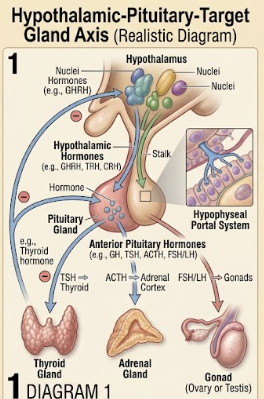
Several endocrine systems are organized into hierarchical axes. In
these pathways, hypothalamic hormones regulate anterior pituitary
hormones, which then stimulate peripheral endocrine glands.
Hypothalamic-pituitary-thyroid axis
TRH
→
TSH
→
Thyroid
→
T3/T4
Hypothalamic-pituitary-adrenal axis
CRH
→
ACTH
→
Adrenal cortex
→
Cortisol
Hypothalamic-pituitary-gonadal axis
GnRH
→
FSH + LH
→
Gonads
→
Sex steroids + inhibin
Growth hormone axis
GHRH
→
GH
→
Liver
→
IGF-1
17
Major Endocrine Disorders: Hyposecretion and Hypersecretion
Endocrine diseases may result from too little hormone secretion,
excessive hormone secretion, abnormal hormone receptors, impaired
transport or failure of the target tissue to respond to a hormone.
Therefore, endocrine disease is not limited to simple hypo- or
hypersecretion.
| Gland/hormone |
Hyposecretion / deficiency |
Hypersecretion / excess |
| GH |
Growth failure in children; altered body composition in adults |
Gigantism in children; acromegaly in adults |
| ADH |
Central diabetes insipidus |
SIADH |
| TSH / thyroid hormones |
Hypothyroidism |
Hyperthyroidism |
| PTH |
Hypocalcemia and tetany |
Hypercalcemia, bone loss and renal stones |
| Cortisol |
Adrenal insufficiency/Addison disease |
Cushing syndrome |
| Aldosterone |
Volume depletion, hyperkalemia |
Primary hyperaldosteronism/Conn syndrome |
| Insulin |
Diabetes mellitus |
Hypoglycemia when insulin activity is excessive relative to glucose availability |
| Testosterone |
Male hypogonadism |
Androgen excess/precocious virilization in selected conditions |
| Ovarian hormones |
Ovarian insufficiency, menstrual disturbance, infertility |
Abnormal steroid excess depending on cause |
Long-answer strategy:
When asked to discuss an endocrine disorder, write:
definition → hormone involved → cause → pathophysiology →
major signs/symptoms → important laboratory/hormonal pattern →
complications.
Acromegaly
Usually results from GH excess after epiphyseal closure.
Enlargement of hands, feet, jaw and facial soft tissues are
characteristic.
Diabetes Insipidus
Reduced ADH secretion or renal resistance to ADH causes excessive
production of dilute urine and marked thirst.
Hypothyroidism
Reduced thyroid hormone action decreases metabolic activity and
may produce fatigue, cold intolerance and weight gain.
Hyperthyroidism
Increased thyroid hormone action increases metabolism and may cause
weight loss, heat intolerance, tremor and tachycardia.
Addison Disease
Primary adrenal insufficiency results in inadequate adrenal cortical
hormone production and can cause hypotension and electrolyte
abnormalities.
Cushing Syndrome
Chronic excess glucocorticoid exposure produces characteristic
metabolic, cardiovascular, skin and musculoskeletal changes.
Diabetes Mellitus
Chronic hyperglycemia occurs because insulin secretion, insulin
action or both are inadequate.
Hyperparathyroidism
Excess PTH can produce hypercalcemia, increased bone turnover and
renal complications.
Pheochromocytoma
Catecholamine-secreting adrenal medullary tumor may produce episodic
hypertension, headache, sweating and palpitations.
19
10-Mark Long Questions with Elaborative Answers
These answers are written in a form suitable for MSc II Semester
theory examination preparation.
Long Question 1
Define the endocrine system. Describe the major endocrine glands,
their hormones and functions.
Answer
The endocrine system is a collection of specialized glands, organs and
tissues that synthesize and secrete hormones. Hormones are chemical
messengers that are transported through the blood or act locally on
nearby cells to regulate specific physiological functions. Endocrine
glands are generally ductless and are closely associated with blood
capillaries. The endocrine system plays a central role in maintenance
of homeostasis, growth, metabolism, reproduction, stress responses,
fluid and electrolyte balance and development.
Major endocrine organs
Important endocrine organs include the hypothalamus, pituitary gland,
pineal gland, thyroid gland, parathyroid glands, thymus, adrenal
glands, pancreatic islets, testes and ovaries. During pregnancy, the
placenta also functions as a temporary endocrine organ.
| Organ |
Major hormone |
Important function |
| Hypothalamus |
TRH, CRH, GnRH, GHRH, somatostatin, dopamine; ADH and oxytocin synthesis |
Regulates pituitary activity and homeostasis |
| Pituitary |
GH, TSH, ACTH, FSH, LH, PRL, ADH, oxytocin |
Controls growth, reproduction, thyroid/adrenal function and water balance |
| Thyroid |
T3, T4, calcitonin |
Metabolism, growth, development and calcium regulation |
| Parathyroid |
PTH |
Maintains calcium and phosphate balance |
| Adrenal cortex |
Aldosterone, cortisol, adrenal androgens |
Fluid balance, metabolism, stress and androgenic effects |
| Adrenal medulla |
Epinephrine, norepinephrine |
Acute stress response |
| Pancreatic islets |
Insulin, glucagon, somatostatin |
Blood glucose and energy regulation |
| Testes |
Testosterone, inhibin B |
Male reproductive development and spermatogenesis |
| Ovaries |
Estrogen, progesterone, inhibin |
Female reproductive function and pregnancy-related changes |
Functions of the endocrine system
-
Growth and development:
GH, thyroid hormones and sex steroids contribute to normal growth
and development.
-
Metabolism:
Thyroid hormones, insulin, glucagon and cortisol regulate energy
utilization and storage.
-
Fluid and electrolyte balance:
ADH and aldosterone regulate water and electrolyte homeostasis.
-
Reproduction:
FSH, LH, estrogen, progesterone and testosterone coordinate
reproductive development and function.
-
Stress response:
Cortisol and adrenal catecholamines help the body respond to
physiological stress.
-
Calcium homeostasis:
PTH, calcitonin and vitamin D-related endocrine pathways regulate
calcium metabolism.
Conclusion
Thus, the endocrine system is a highly integrated regulatory system.
Its hormones act on specific target cells and coordinate multiple
organs to maintain internal stability and adapt the body to changing
physiological demands.
Long Question 2
Describe the pituitary gland, its hormones, functions and disorders
caused by hypo- and hypersecretion.
Answer
The pituitary gland is a small endocrine gland located in the sella
turcica at the base of the brain and connected to the hypothalamus by
the pituitary stalk. It consists of the anterior pituitary and
posterior pituitary. The anterior pituitary synthesizes several
hormones, whereas the posterior pituitary stores and releases ADH and
oxytocin synthesized in the hypothalamus.
Anterior pituitary hormones
Growth hormone promotes body growth and has important metabolic
effects. TSH stimulates the thyroid gland. ACTH stimulates the adrenal
cortex, especially cortisol production. FSH and LH regulate gonadal
function. Prolactin promotes milk production.
Posterior pituitary hormones
ADH promotes water reabsorption by the kidneys, while oxytocin
stimulates uterine contraction and milk ejection.
Major disorders
Growth hormone deficiency during childhood causes impaired growth,
whereas excess GH before epiphyseal closure produces gigantism and
excess GH in adults produces acromegaly.
Excess pituitary ACTH can cause Cushing disease. Deficiency of
pituitary ACTH can cause secondary adrenal insufficiency.
Hyperprolactinemia may cause galactorrhea, menstrual disturbances and
reproductive dysfunction. ADH deficiency causes central diabetes
insipidus, whereas inappropriate excess ADH activity produces SIADH.
Therefore, pituitary disorders can affect multiple organ systems
because pituitary hormones regulate several peripheral endocrine
pathways.
Long Question 3
Describe the thyroid gland, synthesis and functions of thyroid
hormones, and discuss hypo- and hyperthyroidism.
Answer
The thyroid gland is a bilobed endocrine gland located in the anterior
neck. Microscopically, it consists of follicles lined by follicular
cells and containing colloid. Follicular cells synthesize T4 and T3,
whereas parafollicular cells produce calcitonin.
Synthesis of thyroid hormones
Iodide is actively taken up by follicular cells. It is oxidized and
incorporated into tyrosine residues of thyroglobulin. Formation of
MIT and DIT is followed by coupling reactions. DIT plus DIT produces
T4, while MIT plus DIT produces T3. Thyroid hormones are stored in
colloid and released after endocytosis and processing of thyroglobulin.
Functions
Thyroid hormones increase basal metabolic activity and oxygen
consumption. They are essential for normal growth, maturation and
nervous system development. They influence carbohydrate, lipid and
protein metabolism and enhance responsiveness to catecholamines.
Regulation
TRH from the hypothalamus stimulates TSH secretion from the anterior
pituitary. TSH stimulates the thyroid gland to produce T3 and T4.
Increased circulating thyroid hormones exert negative feedback on the
hypothalamus and pituitary.
Hypothyroidism
Hypothyroidism is characterized by inadequate thyroid hormone action.
Patients may develop fatigue, cold intolerance, weight gain,
constipation, dry skin and slowed metabolic activity. In children,
severe untreated hypothyroidism can impair growth and neurological
development.
Hyperthyroidism
Hyperthyroidism produces increased metabolic activity, weight loss,
heat intolerance, sweating, tremor and tachycardia. Graves disease
is an important autoimmune cause and may be associated with
thyroid-associated eye disease.
Long Question 4
Describe the adrenal gland, its hormones, functions and disorders of
hypo- and hypersecretion.
Answer
The adrenal glands are paired endocrine organs situated above the
kidneys. Each adrenal gland consists of an outer cortex and an inner
medulla. The cortex is divided into zona glomerulosa, zona fasciculata
and zona reticularis.
The zona glomerulosa produces aldosterone, the zona fasciculata
produces cortisol and the zona reticularis produces adrenal androgens.
The adrenal medulla produces epinephrine and norepinephrine.
Functions
Aldosterone promotes sodium retention and potassium secretion,
maintaining extracellular fluid volume and blood pressure. Cortisol
regulates metabolism, helps maintain blood glucose during stress and
has anti-inflammatory and immunomodulatory effects. Adrenal androgens
contribute to androgenic activity. Catecholamines mediate rapid
sympathetic responses including increased cardiac activity,
vasoconstriction and energy mobilization.
Hyposecretion
Adrenal insufficiency results in inadequate steroid production.
Primary adrenal insufficiency may cause fatigue, weight loss,
hypotension, electrolyte abnormalities and hyperpigmentation.
Hypersecretion
Excess cortisol produces Cushing syndrome. Excess aldosterone can
produce hypertension and hypokalemia. Catecholamine-secreting
pheochromocytoma can produce episodic hypertension, sweating,
headache and palpitations.
Long Question 5
Describe the endocrine pancreas, its hormones and their functions.
Discuss diabetes mellitus.
Answer
The endocrine pancreas consists mainly of the islets of Langerhans.
These clusters contain alpha, beta, delta and other endocrine cells.
Their hormones regulate glucose and energy metabolism.
Beta cells secrete insulin. Insulin lowers blood glucose by promoting
glucose uptake and storage and by suppressing excessive hepatic
glucose production. Alpha cells secrete glucagon, which increases
blood glucose by stimulating hepatic glycogen breakdown and
gluconeogenesis.
Delta cells produce somatostatin, which inhibits insulin and glucagon
secretion and has additional inhibitory effects on gastrointestinal
functions. Pancreatic polypeptide is secreted by PP cells and
contributes to regulation of pancreatic and gastrointestinal activity.
Diabetes mellitus
Diabetes mellitus is characterized by chronic hyperglycemia caused by
inadequate insulin secretion, impaired insulin action or both.
Type 1 diabetes is usually caused by autoimmune destruction of beta
cells, resulting in marked insulin deficiency. Type 2 diabetes is
characterized primarily by insulin resistance together with progressive
beta-cell dysfunction.
Persistent hyperglycemia can cause microvascular and macrovascular
complications affecting the kidneys, eyes, nerves, heart and blood
vessels. Therefore, pancreatic endocrine dysfunction has consequences
throughout the body.
Long Question 6
Describe the parathyroid gland, functions of parathyroid hormone and
disorders of hypo- and hypersecretion.
Answer
The parathyroid glands are usually four small endocrine glands located
on the posterior surface of the thyroid gland. Their principal
endocrine secretion is parathyroid hormone or PTH.
PTH is released when blood calcium concentration decreases. It acts on
bone and kidney and indirectly on the intestine through stimulation
of calcitriol production. PTH increases renal calcium reabsorption,
decreases phosphate reabsorption and promotes activation of vitamin D.
In bone, PTH increases calcium mobilization indirectly by stimulating
osteoblast-mediated signals that increase osteoclast activity.
Increased calcitriol then promotes intestinal calcium absorption.
Hypoparathyroidism
Deficiency of PTH produces hypocalcemia and increased neuromuscular
excitability. Patients may develop tingling, muscle cramps, tetany,
seizures or cardiac abnormalities when hypocalcemia is severe.
Hyperparathyroidism
Excess PTH produces hypercalcemia and increased bone turnover.
Important manifestations include renal stones, bone pain and skeletal
weakening. Chronic hypercalcemia can also affect kidney function.
Long Question 7
Describe the endocrine functions of testes and ovaries.
Answer
The gonads are reproductive organs that also function as endocrine
organs. The testes produce mainly testosterone and inhibin B, whereas
the ovaries produce estrogens, progesterone, inhibin and AMH.
Testes
LH stimulates Leydig cells to produce testosterone. Testosterone is
essential for male differentiation, development of secondary sexual
characteristics, libido and maintenance of reproductive tissues. FSH
acts mainly on Sertoli cells, which support spermatogenesis and
produce inhibin B. During fetal development, Sertoli cells produce AMH,
which causes regression of Müllerian ducts.
Ovaries
Estrogens promote female reproductive development, secondary sexual
characteristics and endometrial proliferation. Progesterone,
particularly after ovulation, transforms the endometrium into a
secretory state and supports pregnancy. Inhibin suppresses FSH
secretion, while AMH is involved in regulation of follicular
recruitment.
Disorders
Reduced gonadal hormone production can produce hypogonadism,
menstrual disturbances, infertility and altered secondary sexual
development. Excess gonadal steroid production may cause inappropriate
sexual maturation or virilization depending on the hormone and cause.
Long Question 8
Explain the hypothalamic-pituitary axis and endocrine feedback
mechanisms.
Answer
The hypothalamus and pituitary form a central regulatory system for
several endocrine glands. Hypothalamic neurons release regulatory
hormones into the hypophyseal portal circulation, allowing them to
reach the anterior pituitary. These hormones stimulate or inhibit
secretion of anterior pituitary hormones.
The pituitary hormones then act on peripheral endocrine glands such as
the thyroid, adrenal cortex and gonads. The final hormones produced by
these glands generally inhibit further secretion from the hypothalamus
and pituitary through negative feedback.
Examples
-
TRH → TSH → thyroid → T3/T4.
-
CRH → ACTH → adrenal cortex → cortisol.
-
GnRH → FSH/LH → gonads → sex steroids and inhibin.
-
GHRH → GH → IGF-1 and direct tissue effects.
Negative feedback stabilizes endocrine pathways and prevents excessive
hormone production. Positive feedback occurs in selected physiological
situations, such as oxytocin-mediated amplification of uterine
contractions during labour.
Long Question 9
Classify hormones and explain their mechanism of action.
Answer
Hormones can be classified chemically into peptide/protein hormones,
steroid hormones and amine hormones. Their chemical nature determines
their solubility, transport in blood, receptor location and speed of
physiological action.
Peptide and protein hormones
Peptide hormones are generally water-soluble and cannot freely cross
the lipid bilayer. They bind to cell-surface receptors and activate
intracellular second messenger pathways. Examples include insulin,
GH, ADH and PTH.
Steroid hormones
Steroid hormones are lipid-soluble and can cross cell membranes.
They bind intracellular receptors and influence gene transcription.
Examples include cortisol, aldosterone, testosterone, estrogen and
progesterone.
Amine hormones
Amine hormones are derived from amino acids. Catecholamines such as
epinephrine act through membrane receptors, whereas thyroid hormones
act mainly through intracellular nuclear receptors.
Thus, hormone action depends not only on the hormone itself but also on
the receptor expressed by the target cell and the intracellular
signalling machinery available within that cell.
Long Question 10
Discuss major endocrine disorders caused by hormone hyposecretion and
hypersecretion.
Answer
Endocrine disorders occur when hormone secretion or hormone action is
abnormal. Hyposecretion produces inadequate hormone effects, whereas
hypersecretion produces excessive physiological activity. However,
endocrine disease can also arise from receptor defects or abnormal
hormone responsiveness.
| Hormonal abnormality |
Disorder |
Main clinical concept |
| GH deficiency |
Growth hormone deficiency |
Reduced growth in children |
| GH excess |
Gigantism/acromegaly |
Excess growth effects |
| ADH deficiency |
Diabetes insipidus |
Polyuria and polydipsia |
| ADH excess |
SIADH |
Water retention and hyponatremia |
| Thyroid hormone deficiency |
Hypothyroidism |
Reduced metabolic activity |
| Thyroid hormone excess |
Hyperthyroidism |
Increased metabolic activity |
| PTH deficiency |
Hypoparathyroidism |
Hypocalcemia and tetany |
| PTH excess |
Hyperparathyroidism |
Hypercalcemia and increased bone turnover |
| Cortisol deficiency |
Adrenal insufficiency |
Weakness, hypotension and metabolic/electrolyte abnormalities |
| Cortisol excess |
Cushing syndrome |
Chronic glucocorticoid excess |
| Insulin deficiency/action failure |
Diabetes mellitus |
Chronic hyperglycemia |
Understanding the relationship between a hormone and its physiological
action allows the clinical manifestations of endocrine diseases to be
explained rather than memorized as isolated facts.
20
Important 5-Mark Questions
Prepare these as compact but complete answers.
1. Define endocrine gland and differentiate endocrine and exocrine glands.
2. Write the hormones of anterior pituitary and their functions.
3. Describe the hormones of posterior pituitary.
4. Explain the functions of growth hormone.
5. Describe thyroid hormone synthesis.
6. Write a note on thyroid hormones and their functions.
7. Explain the role of parathyroid hormone in calcium homeostasis.
8. Describe the three zones of adrenal cortex.
9. Write a note on adrenal medullary hormones.
10. Explain the hormones of pancreatic islets.
11. Differentiate the actions of insulin and glucagon.
12. Describe the endocrine functions of testes.
13. Describe the endocrine functions of ovaries.
14. Write a short note on melatonin and pineal gland.
15. Explain the endocrine role of thymus.
16. Explain negative feedback in endocrine regulation.
17. Describe hypothalamic-pituitary-thyroid axis.
18. Write a note on Cushing syndrome.
19. Write a note on Addison disease.
20. Explain diabetes mellitus as an endocrine disorder.
21
Important 2-Mark Questions
1. Define hormone.
2. What is an endocrine gland?
3. Name two hormones of the hypothalamus.
4. Name the anterior pituitary hormones.
5. What is the function of GH?
6. What is TSH?
7. Name the posterior pituitary hormones.
8. What is the function of ADH?
9. What is melatonin?
10. Name the thyroid hormones.
11. What is calcitonin?
12. What is PTH?
13. Name the three zones of adrenal cortex.
14. Name two adrenal cortical hormones.
15. Name the adrenal medullary hormones.
16. What is aldosterone?
17. What is cortisol?
18. Name the major pancreatic islet hormones.
19. What is insulin?
20. What is glucagon?
21. Name the major testicular hormone.
22. What is inhibin?
23. Name two ovarian hormones.
24. What is estrogen?
25. What is progesterone?
26. Define hypothyroidism.
27. Define hyperthyroidism.
28. What is acromegaly?
29. What is Addison disease?
30. What is Cushing syndrome?
31. What is diabetes mellitus?
32. What is diabetes insipidus?
33. What is pheochromocytoma?
34. What is negative feedback?
High Yield
Must-Memorize Hormone Tables
Anterior pituitary: hormone → target → function
| Hormone |
Target |
Key function |
| GH |
Many tissues/liver |
Growth and metabolic effects |
| TSH |
Thyroid |
Thyroid hormone synthesis/release |
| ACTH |
Adrenal cortex |
Cortisol production |
| FSH |
Gonads |
Follicular development / spermatogenesis support |
| LH |
Gonads |
Ovulation/corpus luteum and testosterone production |
| PRL |
Mammary gland |
Milk production |
Adrenal cortex: GFR mnemonic
| Zone |
Hormone |
Remember |
| Zona glomerulosa |
Aldosterone |
Salt |
| Zona fasciculata |
Cortisol |
Sugar |
| Zona reticularis |
Adrenal androgens |
Sex |
Very important disorders
| Hormone/gland |
Low activity |
High activity |
| GH |
Growth deficiency |
Gigantism/acromegaly |
| ADH |
Diabetes insipidus |
SIADH |
| Thyroid |
Hypothyroidism |
Hyperthyroidism |
| PTH |
Hypocalcemia/tetany |
Hypercalcemia/bone loss |
| Cortisol |
Addison/adrenal insufficiency |
Cushing syndrome |
| Aldosterone |
Salt wasting/hyperkalemia |
Conn syndrome |
| Insulin |
Diabetes mellitus |
Hypoglycemia |
Diagram Practice
Important Diagrams to Practice for Examination
1. Endocrine System Overview
Draw the major endocrine glands from head to pelvis and label
hypothalamus, pituitary, pineal, thyroid, parathyroids, thymus,
adrenals, pancreas, testes/ovaries.
2. Hypothalamus-Pituitary
Show hypothalamus, pituitary stalk, anterior pituitary and posterior
pituitary with major hormones.
3. Thyroid Histology
Draw thyroid follicles, colloid, follicular cells and
parafollicular cells.
4. Adrenal Cortex
Draw capsule, zona glomerulosa, zona fasciculata, zona reticularis
and medulla.
5. Pancreatic Islet
Show alpha, beta and delta cells and their respective hormones.
6. Endocrine Axes
Practice HPT, HPA and HPG axes with negative feedback.
22
Last-Minute Revision
Revise these points immediately before the examination.
Core Definitions
- Endocrine glands are generally ductless.
- Hormones act on target cells with appropriate receptors.
- Endocrine regulation is essential for homeostasis.
- Negative feedback is the dominant regulatory mechanism.
Pituitary
- GH → growth and metabolism.
- TSH → thyroid.
- ACTH → adrenal cortex.
- FSH/LH → gonads.
- PRL → milk production.
- ADH → water retention.
- Oxytocin → uterine contraction/milk ejection.
Thyroid
- T3 and T4 increase metabolic activity.
- Calcitonin tends to lower plasma calcium.
- TSH stimulates thyroid activity.
- Hypothyroidism → low metabolic activity.
- Hyperthyroidism → high metabolic activity.
Parathyroid
- PTH increases serum calcium.
- PTH decreases renal phosphate reabsorption.
- PTH promotes calcitriol formation.
- Low PTH → hypocalcemia/tetany.
- High PTH → hypercalcemia/bone loss.
Adrenal
- Glomerulosa → aldosterone.
- Fasciculata → cortisol.
- Reticularis → androgens.
- Medulla → epinephrine/norepinephrine.
- Low cortisol → adrenal insufficiency.
- High cortisol → Cushing syndrome.
Pancreas
- Beta → insulin.
- Alpha → glucagon.
- Delta → somatostatin.
- Insulin lowers blood glucose.
- Glucagon raises blood glucose.
- Insulin deficiency/action failure → diabetes mellitus.
Gonads
- Testes → testosterone + inhibin B.
- Leydig cells produce testosterone.
- Sertoli cells support spermatogenesis.
- Ovaries → estrogen + progesterone + inhibin + AMH.
- Estrogen → reproductive development/endometrial proliferation.
- Progesterone → secretory endometrium/pregnancy support.
Must-Know Disorders
- Gigantism
- Acromegaly
- Diabetes insipidus
- SIADH
- Hypothyroidism
- Hyperthyroidism
- Hypoparathyroidism
- Hyperparathyroidism
- Addison disease
- Cushing syndrome
- Conn syndrome
- Diabetes mellitus
- Pheochromocytoma
Exam Strategy
How to Write an Endocrine Long Answer
For a 10-mark endocrine question, avoid writing only a table of
hormones. Begin with a short definition and anatomical introduction,
followed by the structural organization of the gland. Then describe
each hormone in a logical sequence: source → regulation →
target → physiological function → disorder.
For example, if the question asks about the adrenal gland, first
introduce the adrenal gland and divide it into cortex and medulla.
Then discuss the three cortical zones and their hormones. Explain
aldosterone, cortisol and adrenal androgens separately. Then discuss
catecholamines of the medulla. Finally, add adrenal insufficiency,
Cushing syndrome, hyperaldosteronism and pheochromocytoma.
Best 10-mark structure:
- Definition
- Location/gross structure
- Microscopic/basic organization
- Hormones/secretions
- Functions of individual hormones
- Regulation and feedback
- Hyposecretion disorders
- Hypersecretion disorders
- Clinical significance
- Short conclusion
Final Summary
Chapter Summary
The endocrine system is a major regulatory system responsible for
coordination of physiological processes through hormones. The
hypothalamus regulates the pituitary, while the pituitary controls
several peripheral endocrine glands. The thyroid regulates metabolic
activity and development, whereas the parathyroids maintain calcium
homeostasis. The adrenal glands regulate electrolyte balance,
metabolism and stress responses. The pancreatic islets regulate blood
glucose, while the gonads regulate reproductive development and
reproductive physiology.
Endocrine physiology depends heavily on feedback mechanisms, especially
negative feedback. Disturbances may occur because of hormone
hyposecretion, hypersecretion, abnormal receptors or impaired target
tissue responses. Important disorders include acromegaly, diabetes
insipidus, hypothyroidism, hyperthyroidism, hypoparathyroidism,
hyperparathyroidism, Addison disease, Cushing syndrome,
hyperaldosteronism and diabetes mellitus.
For examination preparation, the most useful approach is to connect
each gland with its hormones, targets, physiological actions and
associated disorders. This approach provides both conceptual
understanding and a strong framework for writing detailed MSc-level
long answers.