Table of Contents
1. Introduction to Sensory Organs
The human body continuously receives information from its surroundings through specialized sensory receptors. Light entering the eye, sound waves reaching the ear, odor-producing molecules entering the nasal cavity and chemicals dissolved in saliva stimulating taste receptors on the tongue are all examples of external stimuli. These stimuli are detected by receptor cells and converted into nerve signals.
Sensory organs therefore form an important connection between the environment and the nervous system. They do not simply detect stimuli; their receptor cells are highly specialized to respond to particular forms of energy. Photoreceptors of the retina respond to light, mechanoreceptors in the cochlea respond to mechanical vibrations produced by sound, olfactory receptor neurons respond to odorant molecules, and taste receptor cells respond to chemicals dissolved in saliva.
Although the eye, ear, nose and tongue are traditionally considered the major special sensory organs, each organ represents a complex anatomical and physiological system. Normal sensation requires appropriate receptors, intact sensory nerves, functioning pathways within the central nervous system and appropriate processing within the brain.
2. Classification of Sensory Organs
Sensory receptors can be classified according to the type of stimulus they detect. This classification helps explain why different sensory organs have different anatomical structures and receptor mechanisms.
| Receptor Type | Stimulus Detected | Major Example |
|---|---|---|
| Photoreceptors | Light | Rods and cones of retina |
| Mechanoreceptors | Mechanical deformation, vibration or movement | Organ of Corti and vestibular receptors |
| Chemoreceptors | Chemical substances | Olfactory receptors and taste receptors |
| Thermoreceptors | Temperature changes | Temperature receptors in skin and hypothalamus |
| Nociceptors | Potentially damaging stimuli | Free nerve endings |
Major Special Sensory Organs
| Organ | Major Sense | Principal Receptor | Major Nerve |
|---|---|---|---|
| Eye | Vision | Rods and cones | Optic nerve (CN II) |
| Ear | Hearing and balance | Hair cells | Vestibulocochlear nerve (CN VIII) |
| Nose | Smell | Olfactory receptor neurons | Olfactory nerve (CN I) |
| Tongue | Taste | Taste receptor cells | CN VII, IX and X |
3. Eye
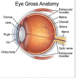
The eye is the specialized sensory organ responsible for vision. It detects light, forms an optical image on the retina and converts light energy into electrical signals that are transmitted through the optic nerve to the brain.
The eye is not simply a hollow optical chamber. It contains transparent refractive media, a vascular and protective wall, specialized neural tissue and several accessory structures that maintain its position, protection, lubrication and movement.
Functions of the Eye
- Detection of visible light.
- Formation of a focused image on the retina.
- Conversion of light energy into electrical signals.
- Transmission of visual information to the brain.
- Accommodation of the lens for near and distant vision.
- Regulation of the amount of light entering the eye through the iris and pupil.
- Contribution to depth perception and color discrimination.
- Participation in visual reflexes such as the pupillary light reflex.
4. Gross Anatomy of the Eye
The eyeball is approximately spherical, with an average adult diameter of about 24 mm. It lies within the bony orbit and is surrounded by orbital fat, muscles, nerves and blood vessels. The anterior portion of the eyeball is relatively exposed and is protected by the eyelids and conjunctiva.
4.1 Coats of the Eyeball
The wall of the eyeball consists of three major coats: an outer fibrous coat, a middle vascular coat and an inner nervous coat. Each coat has a characteristic structure and function.
| Coat | Main Components | Major Functions |
|---|---|---|
| Fibrous coat | Sclera and cornea | Protection, maintenance of shape and refraction |
| Vascular coat / Uvea | Choroid, ciliary body and iris | Nutrition, accommodation and control of light entry |
| Nervous coat | Retina | Detection and initial processing of light |
4.2 Fibrous Coat
Sclera
The sclera is the tough, opaque posterior part of the fibrous coat. It is composed mainly of dense connective tissue rich in collagen fibers. It maintains the shape of the eyeball and provides attachment for the extraocular muscles.
The sclera also provides mechanical protection to the delicate internal structures of the eye. Posteriorly, it is continuous with the dura surrounding the optic nerve.
Cornea
The cornea forms the transparent anterior portion of the fibrous coat. It is highly specialized for transmission and refraction of light. Unlike most tissues of the body, the normal cornea is avascular and receives nutrients mainly by diffusion from the surrounding tissues and aqueous humor.
The cornea has several layers. From anterior to posterior these include corneal epithelium, Bowman's layer, stroma, Descemet's membrane and endothelium. The highly organized arrangement of stromal collagen fibers contributes to corneal transparency.
4.3 Vascular Coat or Uvea
Choroid
The choroid is a highly vascular and pigmented layer situated between the sclera and retina. Its blood vessels supply the outer retina, especially the photoreceptor region. Melanin within the choroid absorbs excess light and helps reduce internal reflection within the eyeball.
Ciliary Body
The ciliary body is a thickened part of the uvea located between the choroid and iris. It contains the ciliary muscle and ciliary processes. The ciliary processes participate in aqueous humor production and provide attachment for the suspensory ligaments of the lens.
Contraction and relaxation of the ciliary muscle alter the tension on the zonular fibers surrounding the lens. This changes the shape of the lens during accommodation.
Iris
The iris is a thin, pigmented diaphragm located anterior to the lens. Its central opening is the pupil. The amount of light entering the eye is regulated by contraction and relaxation of the muscles within the iris.
The sphincter pupillae constricts the pupil, whereas the dilator pupillae enlarges it. Parasympathetic activity generally produces constriction, while sympathetic activity contributes to dilation.
4.4 Lens
The lens is a transparent, biconvex, avascular structure located behind the iris. Its major function is to fine-tune the focusing of light onto the retina. Because the lens is flexible, its curvature can be altered during accommodation.
The lens is enclosed by a capsule and contains lens fibers. Mature lens fibers lose their nuclei and most organelles, allowing light to pass through with minimal scattering.
4.5 Chambers and Ocular Fluids
The anterior part of the eye contains aqueous humor. The posterior cavity contains vitreous body. These transparent media contribute to the optical properties and internal pressure of the eye.
| Structure | Location | Major Function |
|---|---|---|
| Anterior chamber | Between cornea and iris | Contains aqueous humor |
| Posterior chamber | Between iris and lens/ciliary body | Contains aqueous humor |
| Vitreous cavity | Behind lens and extending to retina | Contains vitreous body and supports globe shape |
4.6 Aqueous Humor
Aqueous humor is a clear fluid continuously produced by the ciliary processes. It passes from the posterior chamber through the pupil into the anterior chamber and is mainly drained through the trabecular meshwork and iridocorneal angle into the canal of Schlemm and then into the venous circulation.
Aqueous humor provides nutrients to avascular structures such as the cornea and lens, removes metabolic waste and contributes to the maintenance of intraocular pressure.
4.7 Vitreous Body
The vitreous body is a transparent gel-like substance occupying the large space between the lens and retina. It helps maintain the shape of the eyeball and provides mechanical support to the retina.
4.8 Accessory Structures of the Eye
| Accessory Structure | Function |
|---|---|
| Eyelids | Protect the anterior eye and distribute tears. |
| Conjunctiva | Protective mucous membrane covering the anterior sclera and inner eyelids. |
| Lacrimal apparatus | Produces and drains tears; provides lubrication and antimicrobial protection. |
| Extraocular muscles | Move the eyeball in different directions. |
5. Microscopic Anatomy of the Eye
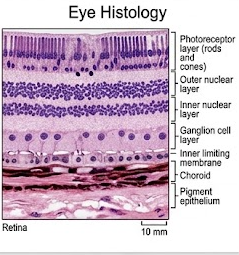
Histologically, the eye contains several specialized tissues. Among them, the retina is particularly important because it contains the neural cells responsible for converting light into electrical signals.
Layers of the Retina
The retina has a complex layered organization. The outer retina is closely associated with the choroid and contains photoreceptors. The inner retina contains bipolar, ganglion and other interneurons. Signals are progressively processed before leaving the eye through the axons of retinal ganglion cells.
| Retinal Component | Major Role |
|---|---|
| Retinal pigment epithelium | Supports photoreceptors, absorbs light and participates in visual pigment recycling. |
| Photoreceptors | Rods and cones detect light. |
| Bipolar cells | Transmit signals from photoreceptors toward ganglion cells. |
| Ganglion cells | Generate action potentials; their axons form the optic nerve. |
| Horizontal and amacrine cells | Provide lateral and integrative processing within the retina. |
6. Retina and Photoreceptors
Rods
Rods are highly sensitive photoreceptors specialized for vision under dim-light conditions. They are especially important for night vision and peripheral vision. Rods contain the visual pigment rhodopsin and are very sensitive to low levels of light.
Rods do not provide high-resolution color vision. Their responses are pooled extensively through retinal circuits, allowing excellent sensitivity but relatively lower spatial resolution.
Cones
Cones function mainly under relatively bright-light conditions and are responsible for high visual acuity and color vision. Three broad classes of cones are sensitive preferentially to different regions of the visible spectrum.
Cone density is particularly high in the foveal region of the macula. The fovea is therefore the region of greatest visual acuity.
| Feature | Rods | Cones |
|---|---|---|
| Light sensitivity | Very high | Lower than rods |
| Major function | Dim-light vision | Color and high-acuity vision |
| Color vision | Absent | Present |
| Distribution | More numerous in peripheral retina | Highly concentrated in foveal region |
| Visual acuity | Lower | High |
Macula and Fovea
The macula lutea is a specialized region of the posterior retina responsible for central detailed vision. At its center lies the fovea centralis, where cone density is particularly high and the overlying retinal layers are displaced, allowing light to reach photoreceptors more directly.
Optic Disc
The optic disc is the point at which retinal ganglion cell axons leave the eyeball to form the optic nerve. Because it lacks photoreceptors, it produces a physiological blind spot in the visual field.
7. Mechanism and Pathway of Vision
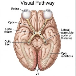
Vision begins when light passes through the transparent optical structures of the eye and reaches the retina. Photoreceptors convert light into changes in membrane potential and neurotransmitter release. Retinal circuits then process these signals before they reach ganglion cells.
Basic Sequence of Visual Processing
Phototransduction
In photoreceptor cells, light activates visual pigments. In rods, the visual pigment is rhodopsin. Light-induced activation of rhodopsin activates a G-protein-mediated cascade involving transducin and phosphodiesterase. This reduces cyclic GMP concentration and causes closure of cGMP-gated sodium channels.
As a result, the photoreceptor membrane becomes hyperpolarized and neurotransmitter release decreases. This change is transmitted through bipolar and ganglion cell pathways and ultimately reaches the visual cortex.
Visual Pathway
Accommodation
Accommodation is the adjustment of lens curvature that allows the eye to focus on objects at different distances. For near vision, parasympathetic stimulation contracts the ciliary muscle. This reduces tension on the zonular fibers, allowing the elastic lens to become more rounded and increase its refractive power.
For distant vision, the ciliary muscle relaxes and zonular tension increases, causing the lens to become flatter. With aging, the lens gradually becomes less elastic, resulting in presbyopia.
Pupillary Light Reflex
The pupillary light reflex is a protective reflex in which exposure of the retina to bright light causes constriction of the pupil. Afferent information travels through the optic nerve and central connections in the midbrain, while efferent parasympathetic signals travel through the oculomotor nerve to the sphincter pupillae.
8. Ear
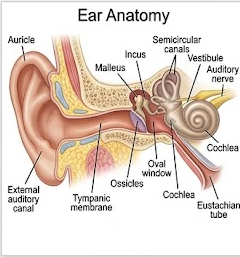
The ear is a specialized sensory organ responsible for two important functions: hearing and maintenance of equilibrium. Anatomically, it is divided into the external ear, middle ear and inner ear.
| Division | Main Structures | Main Function |
|---|---|---|
| External ear | Auricle, external acoustic meatus | Collects and directs sound toward tympanic membrane |
| Middle ear | Tympanic membrane, ossicles, auditory tube | Transmits and amplifies vibrations |
| Inner ear | Cochlea, vestibule, semicircular canals | Hearing and balance |
9. External Ear
Auricle or Pinna
The auricle is the visible external part of the ear. It is composed mainly of elastic cartilage covered by skin. Its irregular contours help collect sound waves and direct them toward the external acoustic meatus.
External Acoustic Meatus
The external acoustic meatus is a canal extending from the auricle toward the tympanic membrane. Its outer part is cartilaginous and its deeper portion is bony. Ceruminous glands produce earwax or cerumen, which contributes to protection of the canal by trapping particles and inhibiting microbial growth.
Tympanic Membrane
The tympanic membrane forms the boundary between the external and middle ear. Sound waves cause the membrane to vibrate. These vibrations are transferred to the ossicles of the middle ear.
10. Middle Ear
The middle ear is an air-filled cavity within the temporal bone. It contains three small auditory ossicles: malleus, incus and stapes. The middle ear transfers vibrations from the tympanic membrane to the inner ear.
Auditory Ossicles
| Ossicle | Position | Function |
|---|---|---|
| Malleus | Attached to tympanic membrane | Receives vibration from tympanic membrane |
| Incus | Between malleus and stapes | Transfers vibration |
| Stapes | Attached to oval window | Transfers vibration into inner ear fluids |
Auditory Tube
The auditory or pharyngotympanic tube connects the middle ear to the nasopharynx. Its major function is to equalize air pressure across the tympanic membrane. Opening of the tube during swallowing or yawning helps maintain pressure balance.
Middle Ear Muscles
The tensor tympani and stapedius muscles modify movement of the auditory ossicles. Their contraction contributes to protection against excessively intense sound by reducing transmission of vibration.
11. Inner Ear
The inner ear is located within the petrous part of the temporal bone and consists of the bony labyrinth and membranous labyrinth. It contains the sensory receptors responsible for hearing and equilibrium.
Bony Labyrinth
The bony labyrinth is a system of fluid-filled cavities containing perilymph. It consists of the cochlea, vestibule and semicircular canals.
Membranous Labyrinth
The membranous labyrinth lies within the bony labyrinth and contains endolymph. It includes the cochlear duct, utricle, saccule and semicircular ducts.
| Structure | Primary Function |
|---|---|
| Cochlea | Hearing |
| Utricle | Detection of linear acceleration and head position, especially in the horizontal plane |
| Saccule | Detection of linear acceleration and head position, especially in the vertical plane |
| Semicircular ducts | Detection of angular/rotational acceleration |
Cochlea
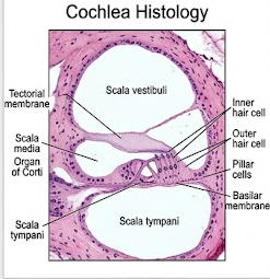
The cochlea is a spiral-shaped structure responsible for hearing. Within the cochlea, the cochlear duct lies between the scala vestibuli and scala tympani. The cochlear duct contains endolymph and houses the organ of Corti.
Organ of Corti
The organ of Corti is located on the basilar membrane within the cochlear duct. It contains inner and outer hair cells supported by specialized supporting cells. The stereocilia of hair cells interact mechanically with the tectorial membrane.
When sound-induced fluid movement causes displacement of the basilar membrane, the hair-cell stereocilia bend. This changes the membrane potential of the hair cells and modifies neurotransmitter release onto sensory nerve fibers of the cochlear division of the vestibulocochlear nerve.
12. Mechanism of Hearing
Hearing begins when sound waves enter the external acoustic meatus and strike the tympanic membrane. The resulting mechanical vibration is transferred through the auditory ossicles to the oval window. Movement of the oval window produces fluid waves within the cochlea.
Role of Basilar Membrane
Different portions of the basilar membrane respond preferentially to different sound frequencies. The basal region is relatively narrow and stiff and responds better to high-frequency sounds, whereas the apical region is wider and more flexible and responds better to low-frequency sounds. This arrangement contributes to frequency discrimination within the cochlea.
Auditory Pathway
Sensory information from cochlear hair cells travels through the cochlear division of the vestibulocochlear nerve to the cochlear nuclei in the brainstem. From there, information passes through several brainstem nuclei and ascends through the lateral lemniscus to the inferior colliculus and medial geniculate body before reaching the primary auditory cortex in the temporal lobe.
13. Vestibular System and Balance
The vestibular apparatus maintains equilibrium by detecting head position and movement. It consists mainly of the utricle, saccule and three semicircular ducts.
Semicircular Ducts
The three semicircular ducts are arranged approximately at right angles to one another. This arrangement allows detection of rotational or angular acceleration in different planes.
Each semicircular duct contains an expanded region called the ampulla. Within the ampulla is the crista ampullaris containing sensory hair cells. Movement of endolymph bends the hair-cell stereocilia and produces receptor signals.
Utricle and Saccule
The utricle and saccule contain sensory regions called maculae. Their hair cells are embedded in a gelatinous membrane containing otoliths composed mainly of calcium carbonate. Gravity and linear acceleration cause movement of the otolithic membrane, stimulating the hair cells.
| Vestibular Structure | Stimulus Detected |
|---|---|
| Semicircular ducts | Angular/rotational acceleration |
| Utricle | Linear acceleration and horizontal head movement |
| Saccule | Linear acceleration and vertical head movement |
Vestibular information is transmitted through the vestibular division of cranial nerve VIII. It contributes to postural control, coordination of eye movements and maintenance of stable vision during head movement.
14. Nose
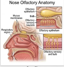
The nose is the first major part of the respiratory tract and also contains the sensory receptors responsible for olfaction. It therefore performs both respiratory and sensory functions.
Major Functions of the Nose
- Provides a passage for inspired and expired air.
- Filters inhaled particles.
- Warms inspired air.
- Humidifies inspired air.
- Provides the organ of smell.
- Contributes to resonance of the voice.
- Participates in defense against inhaled microorganisms and particles.
External Nose
The external nose is formed by bone and cartilage and is covered by skin. It contains the external nares through which air enters and leaves the respiratory tract.
Nasal Cavity
The nasal cavity extends from the nostrils anteriorly to the nasopharynx posteriorly. A nasal septum divides it into right and left cavities. The lateral walls contain superior, middle and inferior nasal conchae, which increase the surface area and produce airflow patterns that promote warming, humidification and filtration.
Respiratory Epithelium
Most of the nasal cavity is lined by pseudostratified ciliated columnar epithelium containing mucus-secreting goblet cells and various supporting cells. Mucus traps inhaled particles, while coordinated ciliary movement helps transport the mucus toward the pharynx.
Olfactory Region
The olfactory epithelium is located mainly in the superior part of the nasal cavity. It contains specialized olfactory receptor neurons, supporting cells and basal cells. The receptor neurons detect odorant molecules and transmit information through the olfactory nerve.
15. Olfaction
Olfaction, or the sense of smell, is a chemical sensory process. Odorant molecules must reach the olfactory epithelium and interact with receptors located on the cilia of olfactory receptor neurons.
Olfactory Receptor Neurons
Olfactory receptor neurons are bipolar neurons. Their dendritic processes extend toward the epithelial surface, while their axons pass through small openings in the cribriform plate of the ethmoid bone to reach the olfactory bulb.
In the olfactory bulb, receptor neuron axons form synapses in structures called glomeruli. The information is then transmitted by mitral and tufted cells through the olfactory tract to different regions of the brain involved in odor perception, emotional responses and memory.
16. Tongue
The tongue is a muscular organ involved in taste, manipulation of food, mastication, swallowing and speech. Its mucosal surface contains specialized structures called papillae, some of which contain taste buds.

Gross Structure of Tongue
The tongue is divided anatomically into an anterior oral part and a posterior pharyngeal part. The two regions are separated by a V-shaped groove called the sulcus terminalis. The anterior two-thirds lies mainly within the oral cavity, while the posterior one-third contributes to the oropharynx.
The tongue is composed primarily of skeletal muscle covered by mucous membrane. The intrinsic muscles alter the shape of the tongue, whereas extrinsic muscles move the tongue as a whole.
Intrinsic Muscles
Intrinsic muscles are located entirely within the tongue. They change the shape of the tongue, allowing it to become shorter, longer, narrower, wider or flattened. These movements are essential for speech and manipulation of food.
Extrinsic Muscles
The major extrinsic muscles are genioglossus, hyoglossus, styloglossus and palatoglossus. They move the tongue in different directions and assist in swallowing and speech.
Tongue Papillae
| Papilla | Location | Characteristics | Taste Buds |
|---|---|---|---|
| Filiform | Most of anterior tongue | Thin, numerous, keratinized; mechanical role | Generally absent |
| Fungiform | Scattered among filiform papillae | Mushroom-shaped | Present |
| Circumvallate | Large papillae arranged near sulcus terminalis | Large and surrounded by a trench | Numerous taste buds on lateral walls |
| Foliate | Posterolateral tongue | Leaf-like folds | Present, particularly prominent in children |
Microscopic Structure of Taste Bud
Taste buds are specialized sensory structures embedded mainly within the epithelium of certain lingual papillae. A taste bud contains receptor cells, supporting cells and basal cells. At its apical end is a small opening called the taste pore through which dissolved chemicals can interact with receptor cell microvilli.
Taste receptor cells are continuously renewed from basal cell populations. When appropriate chemicals stimulate receptor cells, changes in membrane potential lead to neurotransmitter release and activation of sensory nerve fibers.
17. Taste and Gustation
Taste, or gustation, is a chemical sense. Chemicals from food must dissolve in saliva before they can interact with taste receptor cells. Modern physiology recognizes five major taste qualities: sweet, salty, sour, bitter and umami.
| Taste Quality | General Physiological Significance |
|---|---|
| Sweet | Often associated with carbohydrates and energy-rich foods. |
| Salty | Related to detection of ionic substances, especially sodium. |
| Sour | Associated mainly with acidity and hydrogen ion concentration. |
| Bitter | Can provide a protective signal against potentially harmful substances. |
| Umami | Associated particularly with amino acids and certain savory foods. |
Taste Pathway
Taste sensation from the anterior two-thirds of the tongue is carried mainly by the chorda tympani branch of the facial nerve (cranial nerve VII), while taste from the posterior one-third is carried by the glossopharyngeal nerve (cranial nerve IX). Taste from a small region around the epiglottis is carried through the vagus nerve (cranial nerve X).
These sensory fibers enter the brainstem and terminate in the rostral part of the nucleus of the solitary tract. From there, information is relayed through higher centers including the thalamus and ultimately reaches the gustatory cortex, particularly within the insular and adjacent frontal opercular regions.
Tongue Functions Beyond Taste
- Manipulation of food during mastication.
- Formation of a bolus before swallowing.
- Movement of food toward the pharynx.
- Assistance in speech and articulation.
- Contribution to oral cleaning and other movements within the mouth.
18. High-Yield Comparative Tables
Eye vs Ear vs Nose vs Tongue
| Organ | Sense | Receptor | Cranial Nerve | Major Central Destination |
|---|---|---|---|---|
| Eye | Vision | Rods and cones | II | Visual cortex |
| Ear | Hearing | Cochlear hair cells | VIII | Auditory cortex |
| Ear | Balance | Vestibular hair cells | VIII | Vestibular nuclei and cerebellar/cortical systems |
| Nose | Smell | Olfactory receptor neurons | I | Olfactory and limbic-associated regions |
| Tongue | Taste | Taste receptor cells | VII, IX, X | Gustatory cortex |
Major Sensory Receptors
| Receptor | Location | Stimulus |
|---|---|---|
| Rods | Retina | Low-intensity light |
| Cones | Retina | Light, color and high-resolution vision |
| Cochlear hair cells | Organ of Corti | Sound-induced mechanical vibration |
| Vestibular hair cells | Utricle, saccule and semicircular ducts | Acceleration and head movement |
| Olfactory receptor neurons | Olfactory epithelium | Odorant molecules |
| Taste receptor cells | Taste buds | Dissolved chemicals |
Important Cranial Nerves
| Cranial Nerve | Number | Sensory Role |
|---|---|---|
| Olfactory | CN I | Smell |
| Optic | CN II | Vision |
| Facial | CN VII | Taste from anterior two-thirds of tongue |
| Glossopharyngeal | CN IX | Taste from posterior one-third of tongue |
| Vagus | CN X | Taste from small region near epiglottis |
| Vestibulocochlear | CN VIII | Hearing and equilibrium |
19. Clinical Correlations
Knowledge of the anatomy of sensory organs is essential for understanding common sensory disorders. A lesion may affect the receptor, peripheral nerve, sensory pathway or central processing area. Therefore, the clinical manifestation depends on the exact anatomical site involved.
Cataract
Cataract is an opacity of the normally transparent lens. It interferes with the passage of light and may progressively reduce visual acuity.
Glaucoma
Glaucoma refers to a group of disorders involving optic nerve damage, often associated with abnormal intraocular pressure. Progressive damage can result in visual field loss.
Myopia
In myopia, distant objects are focused in front of the retina when accommodation is relaxed. Concave lenses are commonly used for correction.
Hypermetropia
In hypermetropia, the optical system tends to focus images behind the retina when accommodation is relaxed. Convex lenses can be used for correction.
Presbyopia
Age-related reduction in lens elasticity and accommodative ability makes near vision difficult.
Conjunctivitis
Inflammation of the conjunctiva can produce redness, irritation, discharge and discomfort.
Otitis Media
Infection or inflammation of the middle ear can interfere with normal transmission of sound and may cause pain or conductive hearing impairment.
Conductive Hearing Loss
Occurs when sound transmission through the external or middle ear is impaired, for example by obstruction or middle-ear pathology.
Sensorineural Hearing Loss
Results from damage involving cochlear hair cells, the auditory nerve or higher auditory pathways.
Vertigo
Vertigo is a false sensation of movement or spinning and may result from dysfunction of the vestibular apparatus or its central connections.
Anosmia
Anosmia refers to loss of the sense of smell. It may occur due to nasal obstruction, epithelial injury, olfactory nerve damage or central nervous system disease.
Ageusia
Ageusia means loss of taste. Disturbances may involve taste receptor cells, sensory nerves or central pathways.
20. Important 10-Mark Long Questions
Introduction
The eye is the specialized organ of vision. It receives light from the external environment, focuses the light onto the retina, converts the light stimulus into neural signals and transmits these signals to the brain for interpretation. The eyeball is situated in the orbit and is protected by the eyelids, conjunctiva, lacrimal apparatus and orbital structures.
Gross Structure
The wall of the eyeball consists of three principal coats: fibrous, vascular and nervous. The fibrous coat is formed by the sclera and cornea. The sclera is opaque, strong and protective, while the cornea is transparent and forms an important refractive surface.
The vascular coat, or uvea, consists of the choroid, ciliary body and iris. The choroid is highly vascular and supplies the outer retina. The ciliary body produces aqueous humor and participates in accommodation. The iris regulates the amount of light entering through the pupil.
The nervous coat is the retina. It contains photoreceptors, interneurons and ganglion cells. Rods are mainly responsible for dim-light vision, whereas cones provide high-acuity and color vision.
Lens and Ocular Media
The lens is a transparent biconvex structure situated behind the iris. It changes its curvature during accommodation to focus objects at different distances. Aqueous humor occupies the anterior and posterior chambers, while vitreous body occupies the large cavity behind the lens. These media contribute to refraction and maintenance of the internal environment and shape of the eyeball.
Retina
The retina is the sensory layer of the eye. Light is detected by rods and cones. Signals are processed by bipolar, horizontal, amacrine and ganglion cells. The axons of ganglion cells leave the eye through the optic disc and form the optic nerve.
Visual Pathway
Visual information passes from the retina through the optic nerve to the optic chiasma, where fibers from the nasal retina cross. The fibers then continue in the optic tract to the lateral geniculate nucleus. From there, optic radiations carry information to the primary visual cortex in the occipital lobe.
Functions
- Detection of light.
- Formation of focused retinal images.
- Color discrimination.
- High-resolution central vision.
- Dim-light and peripheral vision.
- Depth and spatial perception.
- Accommodation for near and distant objects.
- Participation in pupillary light reflexes.
Conclusion
Thus, the eye is a highly specialized optical and neural organ. Its transparent refractive structures focus light on the photoreceptors of the retina, while the retinal neural network converts and processes the information before transmitting it to the visual centers of the brain.
Introduction
The ear is a specialized sensory organ responsible for hearing and equilibrium. Anatomically it is divided into external, middle and inner ear. The external and middle ear primarily participate in sound transmission, whereas the inner ear contains the sensory receptors for both hearing and balance.
External Ear
The external ear consists of the auricle and external acoustic meatus. The auricle collects sound waves and directs them toward the external acoustic canal. The sound waves strike the tympanic membrane and cause it to vibrate.
Middle Ear
The middle ear is an air-filled cavity containing three ossicles: malleus, incus and stapes. The malleus receives vibration from the tympanic membrane, the incus transmits the vibration and the stapes transfers it to the oval window of the inner ear. The auditory tube helps equalize pressure between the middle ear and nasopharynx.
Inner Ear
The inner ear contains the cochlea, vestibule and semicircular canals. The cochlea contains the organ of Corti, which is the principal sensory receptor for hearing. The vestibular apparatus consists of the utricle, saccule and semicircular ducts and is responsible for equilibrium.
Mechanism of Hearing
Sound waves enter the external acoustic meatus and vibrate the tympanic membrane. These vibrations are amplified and transmitted by the ossicles to the oval window. Movement of the oval window generates fluid waves in the cochlea. The resulting movement of the basilar membrane bends the stereocilia of cochlear hair cells in the organ of Corti. This produces electrical changes in the receptor cells and alters neurotransmitter release onto sensory nerve fibers.
Auditory information travels through the cochlear division of cranial nerve VIII to the cochlear nuclei and then through ascending auditory pathways to the medial geniculate body and auditory cortex.
Maintenance of Balance
The semicircular ducts detect angular acceleration, while the utricle and saccule detect linear acceleration and head position. Their hair cells are stimulated by movement of endolymph or otolithic membranes. Vestibular information travels through the vestibular nerve and contributes to postural control, eye stabilization and spatial orientation.
Conclusion
Therefore, the ear performs two closely related sensory functions. The cochlear apparatus converts sound vibration into neural information for hearing, while the vestibular apparatus detects movement and position of the head and contributes to maintenance of equilibrium.
Introduction
The nose is an important part of the upper respiratory tract and also serves as the peripheral organ of smell. It consists of an external nose and an internal nasal cavity. Its respiratory and sensory functions are closely related to its specialized epithelial lining.
Structure
The external nose is formed by bone and cartilage and contains the external nares. The nasal cavity is divided into right and left portions by the nasal septum. Its lateral wall contains superior, middle and inferior conchae that increase surface area and create airflow patterns.
Most of the nasal cavity is lined by respiratory epithelium. Mucus secreted by goblet cells traps inhaled particles, while cilia transport the mucus toward the pharynx. The rich blood supply helps warm inspired air, and the moist mucosal surface contributes to humidification.
Olfactory Region
The olfactory epithelium is located mainly in the superior nasal cavity. It contains bipolar olfactory receptor neurons, supporting cells and basal cells. Odorant molecules dissolve in the mucus and interact with receptors on the cilia of olfactory receptor neurons.
Olfactory Pathway
Axons of olfactory receptor neurons pass through the cribriform plate and terminate in the olfactory bulb. Within the bulb, synaptic organization occurs in glomeruli. Mitral and tufted cells then carry information through the olfactory tract to higher brain regions involved in odor identification, emotional processing and memory.
Functions
- Smell and odor discrimination.
- Filtration of inhaled particles.
- Warming of inspired air.
- Humidification of inspired air.
- Defense against inhaled particles and microorganisms.
- Contribution to voice resonance.
Introduction
The tongue is a muscular organ of the oral cavity involved in taste, mastication, swallowing and speech. Its mucosal surface contains several types of papillae, and specialized taste buds detect dissolved chemical substances.
Gross Structure
The tongue is divided into an anterior two-thirds and posterior one-third by the sulcus terminalis. It consists mainly of skeletal muscle covered by mucosa. Intrinsic muscles change the shape of the tongue, whereas extrinsic muscles move it within the oral cavity.
Papillae
Four major types of papillae are recognized: filiform, fungiform, circumvallate and foliate. Filiform papillae are numerous and mainly perform a mechanical function. Fungiform, circumvallate and foliate papillae contain taste buds.
Taste Buds
Taste buds contain receptor cells, supporting cells and basal cells. Their apical surface opens through a taste pore. Chemicals dissolved in saliva enter the taste pore and interact with receptor mechanisms on the taste receptor cells.
Taste Sensation
Taste receptor cells detect five major taste qualities: sweet, salty, sour, bitter and umami. The receptor cells respond to appropriate chemical stimuli by changing their membrane potential and releasing signaling molecules onto sensory nerve fibers.
Taste Pathway
Taste from the anterior two-thirds of the tongue is transmitted mainly through the facial nerve, while taste from the posterior one-third is transmitted through the glossopharyngeal nerve. Taste from a small region near the epiglottis travels through the vagus nerve. These signals reach the brainstem, are relayed through higher sensory pathways and ultimately reach the gustatory cortex.
Functions
- Taste sensation.
- Food manipulation.
- Formation of food bolus.
- Assistance in swallowing.
- Speech and articulation.
Introduction
Special sensory organs are specialized anatomical structures that detect particular environmental stimuli and transmit information to the central nervous system. The major special sensory organs covered in this chapter are the eye, ear, nose and tongue.
| Organ | Sensory Function | Receptor | Major Nerve |
|---|---|---|---|
| Eye | Vision | Rods and cones | Optic nerve |
| Ear | Hearing and balance | Hair cells | Vestibulocochlear nerve |
| Nose | Smell | Olfactory receptor neurons | Olfactory nerve |
| Tongue | Taste | Taste receptor cells | Facial, glossopharyngeal and vagus nerves |
The eye detects light and forms an optical image on the retina. The retina contains rods and cones that convert light into neural signals. The ear detects sound through the cochlea and maintains equilibrium through the vestibular apparatus. The nose contains olfactory receptor neurons that detect odorant molecules, while the tongue contains taste buds that detect dissolved chemical stimuli.
Thus, although these organs differ markedly in structure, they follow a common physiological principle: an appropriate stimulus activates a specialized receptor, the receptor converts the stimulus into a neural signal, and sensory pathways transmit the information to the central nervous system for interpretation.
Retina
The retina is the sensory neural layer of the eye. It contains photoreceptors, interneurons and ganglion cells arranged in a highly organized manner. Rods and cones are located toward the outer retinal region, close to the retinal pigment epithelium. Their signals are transmitted to bipolar cells and subsequently to ganglion cells. Horizontal and amacrine cells contribute to lateral and integrative processing.
Ganglion cell axons converge at the optic disc and form the optic nerve. The foveal region contains a high density of cones and specialized retinal organization that permits high visual acuity.
Organ of Corti
The organ of Corti lies on the basilar membrane within the cochlear duct. It contains sensory inner and outer hair cells and supporting cells. Hair cells possess stereocilia that respond to mechanical displacement produced by movement of the basilar membrane.
Bending of stereocilia alters ion conductance and changes the membrane potential of the hair cells. Neurotransmitter release then activates sensory fibers of the cochlear nerve. Different regions of the basilar membrane respond preferentially to different frequencies, allowing the auditory system to analyze sound frequency.
Conclusion
The microscopic organization of both organs is directly related to their sensory functions. The retina is organized for phototransduction and neural processing of visual information, whereas the organ of Corti is specialized for mechanical transduction of sound vibrations.
21. Important 5-Mark Questions
1. Write a short note on rods and cones.
Rods and cones are photoreceptors of the retina. Rods are highly sensitive to dim light and are important for night and peripheral vision but do not provide color discrimination. Cones function mainly in bright light and are responsible for color vision and high visual acuity. Cones are particularly concentrated in the foveal region.
2. Describe the three coats of the eyeball.
The eyeball has three coats. The outer fibrous coat consists of sclera and cornea and provides protection and refraction. The middle vascular coat consists of choroid, ciliary body and iris and provides nutrition, accommodation and control of light entry. The inner nervous coat is the retina, which contains photoreceptors and neural cells involved in vision.
3. Explain accommodation of the eye.
Accommodation is the adjustment of lens curvature for focusing objects at different distances. During near vision, contraction of the ciliary muscle reduces zonular tension and allows the lens to become more rounded, increasing its refractive power. During distant vision, the ciliary muscle relaxes and the lens becomes flatter.
4. Describe the auditory ossicles.
The three auditory ossicles are malleus, incus and stapes. The malleus is attached to the tympanic membrane, the incus lies between malleus and stapes, and the stapes contacts the oval window. They transmit and amplify vibrations from the tympanic membrane to the inner ear.
5. Write a note on organ of Corti.
The organ of Corti is the principal sensory receptor for hearing. It lies on the basilar membrane within the cochlear duct and contains inner and outer hair cells and supporting cells. Movement of the basilar membrane bends the stereocilia of hair cells, producing receptor potentials and neurotransmitter release to auditory nerve fibers.
6. Explain the vestibular apparatus.
The vestibular apparatus consists mainly of the semicircular ducts, utricle and saccule. Semicircular ducts detect angular acceleration, whereas utricle and saccule detect linear acceleration and head position. Hair cells within these structures convert mechanical movement into neural signals carried by the vestibular division of cranial nerve VIII.
7. Describe the olfactory epithelium.
Olfactory epithelium is located mainly in the superior nasal cavity. It contains bipolar olfactory receptor neurons, supporting cells and basal cells. Odorant molecules interact with receptors on olfactory neuron cilia. Axons of these neurons pass through the cribriform plate to the olfactory bulb.
8. Describe tongue papillae.
The four major papillae are filiform, fungiform, circumvallate and foliate. Filiform papillae are numerous and mainly mechanical. Fungiform, circumvallate and foliate papillae contain taste buds. Circumvallate papillae are large and arranged near the sulcus terminalis.
9. Write a note on taste pathway.
Taste from the anterior two-thirds of the tongue travels mainly through the facial nerve, while the posterior one-third travels through the glossopharyngeal nerve. A small region near the epiglottis is supplied through the vagus nerve. The information enters the brainstem, is relayed through higher sensory pathways and reaches the gustatory cortex.
10. Differentiate conductive and sensorineural hearing loss.
| Conductive Hearing Loss | Sensorineural Hearing Loss |
|---|---|
| External or middle ear involvement | Inner ear, auditory nerve or central pathway involvement |
| Sound transmission is impaired | Sound transduction or neural processing is impaired |
| Examples include canal obstruction and middle-ear disease | Examples include cochlear hair-cell or auditory nerve damage |
22. Important 2-Mark Questions
| Question | Expected Answer |
|---|---|
| Define sensory organ. | An organ containing specialized receptors that detect stimuli and transmit sensory information to the nervous system. |
| Name the three coats of the eyeball. | Fibrous coat, vascular coat and nervous coat. |
| Name the two photoreceptors of retina. | Rods and cones. |
| What is the blind spot? | The optic disc, where the optic nerve leaves the eye and photoreceptors are absent. |
| What is accommodation? | Adjustment of lens curvature to focus images of objects at different distances on the retina. |
| What is the function of iris? | It regulates the amount of light entering the eye by controlling pupil diameter. |
| Name the three auditory ossicles. | Malleus, incus and stapes. |
| What is the organ of Corti? | The principal sensory receptor organ for hearing located within the cochlear duct. |
| Which cranial nerve carries hearing? | Cochlear division of cranial nerve VIII. |
| Which structures maintain balance? | Semicircular ducts, utricle and saccule. |
| What is the function of semicircular ducts? | They detect angular or rotational acceleration of the head. |
| What is olfaction? | The sense of smell produced by detection of odorant molecules by olfactory receptors. |
| Which cranial nerve carries smell? | Olfactory nerve, cranial nerve I. |
| Name the four major types of tongue papillae. | Filiform, fungiform, circumvallate and foliate. |
| Which papillae generally lack taste buds? | Filiform papillae. |
| Name the five basic taste qualities. | Sweet, salty, sour, bitter and umami. |
| Which nerve carries taste from anterior two-thirds of tongue? | Facial nerve, mainly through the chorda tympani. |
| Which nerve carries taste from posterior one-third? | Glossopharyngeal nerve, cranial nerve IX. |
| Define cataract. | Opacity of the normally transparent lens of the eye. |
| Define glaucoma. | A group of disorders involving progressive optic nerve damage, often associated with abnormal intraocular pressure. |
| What is presbyopia? | Age-related reduction in accommodation causing difficulty in near vision. |
| Define anosmia. | Loss or absence of the sense of smell. |
| Define ageusia. | Loss or marked reduction of taste sensation. |
23. Diagrams You Should Practice
Eye
- Gross structure of eye
- Three coats of eyeball
- Retinal layers
- Visual pathway
- Accommodation mechanism
Ear
- External, middle and inner ear
- Auditory ossicles
- Cochlea
- Organ of Corti
- Vestibular apparatus
- Auditory pathway
Nose & Tongue
- Nasal cavity
- Olfactory epithelium
- Olfactory pathway
- Tongue papillae
- Taste bud
- Taste pathway
24. High-Yield Examination Points
- Eye: Three coats are fibrous, vascular and nervous.
- Cornea: Transparent anterior part of fibrous coat and an important refractive surface.
- Iris: Controls pupil diameter and therefore regulates light entry.
- Retina: Contains rods and cones and performs phototransduction.
- Rods: Important for dim-light vision.
- Cones: Important for color and high-acuity vision.
- Fovea: Region of greatest visual acuity.
- Optic disc: Physiological blind spot.
- Ear: Responsible for hearing and equilibrium.
- Ossicles: Malleus, incus and stapes.
- Organ of Corti: Principal receptor organ for hearing.
- Semicircular ducts: Detect angular acceleration.
- Utricle and saccule: Detect linear acceleration and head position.
- Nose: Performs respiratory and olfactory functions.
- Olfactory receptor neurons: Specialized bipolar neurons in olfactory epithelium.
- Tongue: Performs taste, food manipulation, swallowing and speech functions.
- Filiform papillae: Mainly mechanical and generally lack taste buds.
- Circumvallate papillae: Large papillae located near the sulcus terminalis and contain many taste buds.
- Anterior two-thirds taste: Mainly facial nerve through chorda tympani.
- Posterior one-third taste: Glossopharyngeal nerve.
25. Last-Minute Revision
Eye — Remember This Chain
Cornea → aqueous humor → pupil → lens → vitreous body → retina → optic nerve → optic chiasma → optic tract → lateral geniculate nucleus → optic radiation → visual cortex.
Ear — Hearing Chain
Auricle → external acoustic meatus → tympanic membrane → malleus → incus → stapes → oval window → cochlea → organ of Corti → cochlear nerve → auditory pathway → auditory cortex.
Ear — Balance
Semicircular ducts → angular acceleration. Utricle → mainly horizontal linear acceleration. Saccule → mainly vertical linear acceleration.
Nose — Smell Chain
Odorant → olfactory epithelium → olfactory receptor neuron → cribriform plate → olfactory bulb → olfactory tract → higher olfactory brain regions.
Tongue — Taste Chain
Chemical stimulus → taste bud → CN VII / IX / X → nucleus of solitary tract → higher relay → gustatory cortex.
Four Most Important 10-Mark Topics
- Structure and functions of the eye.
- Structure of ear with mechanism of hearing and balance.
- Structure and functions of nose with olfaction.
- Structure and functions of tongue with taste sensation.
Four Diagrams to Practice Before Examination
- Well-labelled diagram of the eye.
- Well-labelled diagram of the ear.
- Visual pathway.
- Cochlea and organ of Corti.
- Nasal cavity and olfactory epithelium.
- Tongue showing papillae and taste bud.
26. Chapter Summary
The sensory organs provide specialized mechanisms for detecting important changes in the external environment. The eye is a complex optical and neural organ that detects light through rods and cones and transmits visual information to the brain through the optic pathway. The ear contains the cochlea for hearing and the vestibular apparatus for equilibrium. The nose contains olfactory receptor neurons that detect odorant molecules and transmit information to the olfactory bulb and higher brain regions. The tongue contains taste buds that detect dissolved chemical stimuli and also performs important mechanical functions in eating, swallowing and speech.
In all four sensory systems, the basic principle is similar: an appropriate stimulus interacts with a specialized receptor, receptor transduction produces a change in electrical activity, sensory pathways transmit the information toward the central nervous system and the brain interprets the signal as a particular sensory experience. Understanding this common principle while learning the unique anatomy of each organ is the key to mastering the sensory organs chapter.