MB 551 Human Anatomy & Physiology
• 4 Hours • MSc Medical Microbiology Semester II
Detailed study of the male and female reproductive organs,
their gross and microscopic anatomy, functions, reproductive
physiology and important clinical correlations.
Exam Focus:
Reproductive System is a broad 4-hour chapter. For long questions,
do not write only the names of organs. A complete answer should include
the introduction, classification of organs, gross anatomy,
microscopic anatomy, functions and relevant hormonal/reproductive roles.
The male reproductive system is commonly organized around the testes,
ducts, accessory glands and penis, while the female system is organized
around the ovaries, uterine tubes, uterus, vagina and external genitalia.
The reproductive system is the group of organs responsible for reproduction,
production of gametes, secretion of reproductive hormones, fertilization and,
in females, support of pregnancy, fetal development and childbirth. Unlike
many other body systems, the reproductive system is not primarily necessary
for the survival of an individual, but it is essential for continuation of
the species.
The reproductive system consists of both primary reproductive organs
(gonads) and accessory reproductive organs. The male
gonads are the testes, whereas the female gonads are the ovaries. The gonads
perform two major functions: they produce reproductive cells or gametes and
secrete reproductive hormones. The testes produce spermatozoa and mainly
secrete testosterone, while the ovaries produce ova and secrete estrogen and
progesterone.
The accessory reproductive organs provide pathways for transport, maturation
and delivery of gametes. In females, the reproductive tract also provides the
site for fertilization, implantation, pregnancy and parturition. Therefore,
the reproductive system has both anatomical and endocrine functions.
Important concept:
The reproductive system is closely integrated with the endocrine system.
The hypothalamus, pituitary gland and gonads form the
hypothalamic-pituitary-gonadal (HPG) axis, which regulates
gametogenesis and secretion of reproductive hormones.
2. Major Reproductive Organs
System
Primary Organ/Gonad
Major Accessory Organs
Main Hormones
Male
Testes
Epididymis, vas deferens, ejaculatory ducts, urethra,
seminal vesicles, prostate, bulbourethral glands and penis
Testosterone, inhibin B
Female
Ovaries
Uterine tubes, uterus, cervix, vagina and external genitalia
Estrogen, progesterone, inhibin, AMH
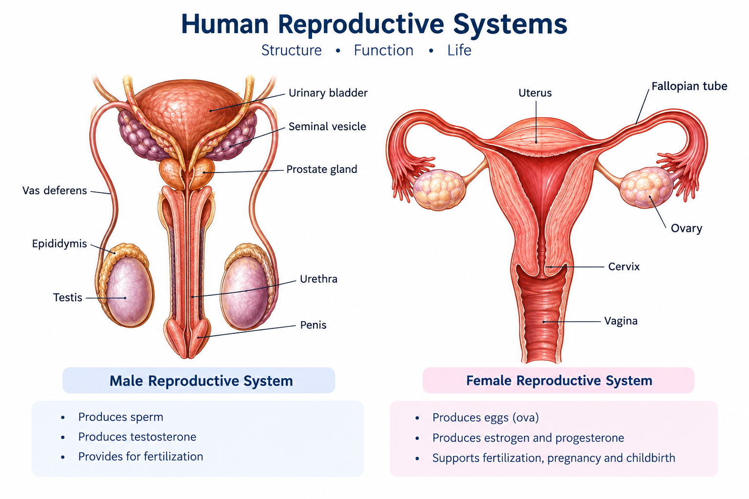
Figure: Anatomical overview of the male and female reproductive systems.
3. Male Reproductive System
The male reproductive system consists of the testes, a system of ducts,
accessory reproductive glands and external genitalia. The testes are the
primary male reproductive organs and are responsible for sperm production
and secretion of testosterone.
The sperm produced within the seminiferous tubules pass through a series of
ducts before reaching the exterior. The epididymis provides an important site
for sperm maturation and storage. The vas deferens transports sperm toward
the pelvic cavity. The seminal vesicles, prostate and bulbourethral glands
contribute secretions that combine with sperm to form semen.
The penis acts as the organ of copulation and provides a passage for semen
during ejaculation. The male urethra serves as a common passage for urine
and semen, although the two are not normally expelled simultaneously.
Primary Gonad
Testis — sperm production and testosterone secretion.
Sperm Maturation
Epididymis — maturation, concentration and storage of sperm.
Sperm Transport
Vas deferens and ejaculatory ducts transport sperm.
Accessory Secretions
Seminal vesicles, prostate and bulbourethral glands contribute seminal fluid.
4. Testis
Location and Gross Anatomy
The testes are paired male gonads located within the scrotum. The scrotal
position keeps the testes at a temperature slightly below core body
temperature, which is important for normal spermatogenesis.
Each testis is approximately oval in shape and is covered by several
protective layers. The outer covering includes the tunica vaginalis and a
dense connective tissue capsule called the tunica albuginea.
The tunica albuginea sends connective tissue septa into the testis and
divides it into numerous testicular lobules.
Each lobule contains one or more highly coiled seminiferous tubules.
These tubules converge toward the mediastinum testis and ultimately connect
with the duct system that carries sperm to the epididymis.
Microscopic Anatomy of Testis
The testis is composed mainly of seminiferous tubules and interstitial tissue.
The seminiferous tubules are lined by a specialized germinal epithelium
containing developing germ cells and Sertoli cells.
Between the seminiferous tubules is interstitial connective tissue containing
the Leydig cells. These endocrine cells produce testosterone
under the influence of luteinizing hormone (LH).
Figure: Gross organization and microscopic structure of the testis,
including seminiferous tubules and interstitial Leydig cells.
Important Cells of Testis
Cell
Location
Main Function
Spermatogenic cells
Seminiferous epithelium
Develop into mature spermatozoa.
Sertoli cells
Seminiferous tubules
Support developing germ cells, form the blood-testis barrier,
secrete inhibin B and produce androgen-binding protein.
Leydig cells
Interstitial tissue
Secrete testosterone.
Functions of Testis
Production of spermatozoa.
Secretion of testosterone.
Secretion of inhibin B by Sertoli cells.
Support of male reproductive development.
Maintenance of male secondary sexual characteristics.
Support of libido and anabolic processes.
5. Seminiferous Tubules and Spermatogenesis
Seminiferous tubules are the functional sites of sperm production within the
testis. Their walls contain a specialized stratified germinal epithelium.
Spermatogenic cells are arranged from the basement membrane toward the lumen
according to their stage of development.
Sertoli cells extend from the basement membrane toward the lumen and provide
nutritional and structural support to developing germ cells. Tight junctions
between Sertoli cells contribute to the blood-testis barrier, which separates
developing germ cells from many components of systemic circulation.
Stages of Spermatogenesis
Spermatogonia: diploid stem/germ cells located near the basement membrane.
Primary spermatocytes: undergo the first meiotic division.
Secondary spermatocytes: produced after meiosis I and rapidly undergo meiosis II.
Spermatids: haploid cells formed after meiosis II.
Spermatozoa: mature sperm cells produced through spermiogenesis.
Spermiogenesis is the transformation of spermatids into structurally
specialized spermatozoa. During this process, the nucleus becomes condensed,
the acrosome develops, excess cytoplasm is removed and the flagellum develops.
The resulting spermatozoa are released into the lumen of the seminiferous
tubules.
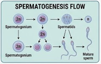
Figure: Sequential stages of spermatogenesis from spermatogonium to
mature spermatozoon.
Sperm Structure
Part
Main Features
Function
Head
Nucleus and acrosome
Contains paternal genetic material; acrosome contains enzymes involved in fertilization.
Neck
Connects head and middle piece
Structural connection.
Middle piece
Contains numerous mitochondria
Provides ATP for sperm movement.
Tail
Flagellum
Produces motility.
6. Epididymis
The epididymis is a long, highly coiled duct attached to the posterior aspect
of each testis. Anatomically it is divided into the head, body and tail.
The head receives sperm from the efferent ductules, while the tail continues
into the vas deferens.
Microscopic Anatomy
The epididymis is lined mainly by pseudostratified columnar epithelium with
stereocilia. The stereocilia are long non-motile microvilli that increase
surface area and participate in absorption and modification of luminal fluid.
Smooth muscle surrounds the duct and contributes to movement of sperm.
Functions
Sperm maturation.
Acquisition of progressive motility.
Concentration of sperm.
Storage of sperm, especially in the tail.
Transport of sperm toward the vas deferens.
Clinical correlation:
Inflammation of the epididymis is called epididymitis.
It may be associated with infection and can produce scrotal pain and
swelling.
7. Vas Deferens
The vas deferens, or ductus deferens, is a thick muscular tube that carries
sperm from the epididymis toward the ejaculatory duct. It passes through the
spermatic cord and inguinal canal before entering the pelvic cavity.
Microscopic Anatomy
The wall of the vas deferens consists of a mucosa lined by pseudostratified
columnar epithelium, a thick muscular layer and an outer connective tissue
layer. The prominent smooth muscle allows powerful peristaltic contractions
during ejaculation.
Function
Its principal function is rapid transport of sperm from the epididymis toward
the ejaculatory duct during ejaculation. The thick muscular wall is particularly
important because sperm must be transported efficiently over a relatively
long distance.
8. Ejaculatory Duct and Male Urethra
Ejaculatory Duct
Each ejaculatory duct is formed by the union of the duct of the seminal
vesicle with the ampulla of the vas deferens. It passes through the prostate
and opens into the prostatic urethra.
The ejaculatory ducts therefore provide a short final pathway for the mixture
of sperm and seminal vesicle secretion before it enters the urethra.
Male Urethra
The male urethra is a common passageway for urine and semen. It extends from
the urinary bladder through the prostate and penis.
Part
Location
Important Feature
Prostatic urethra
Within prostate
Receives ejaculatory ducts and prostatic ducts.
Membranous urethra
Through external urethral sphincter region
Short and surrounded by skeletal muscle.
Spongy urethra
Within penis
Longest portion; surrounded by corpus spongiosum.
9. Seminal Vesicles
The seminal vesicles are paired accessory reproductive glands located
posterior to the urinary bladder. Their ducts join the vas deferens to form
the ejaculatory ducts.
The secretion of the seminal vesicles contributes a substantial portion of
seminal plasma. It is rich in fructose, which provides an energy source for
sperm. The secretion also contains substances that support sperm function
and contribute to the appropriate environment for sperm transport.
Microscopic Structure
The seminal vesicle has a highly folded mucosa, producing an irregular
luminal appearance. It is lined mainly by pseudostratified columnar
epithelium and contains a muscular wall that helps expel its secretion
during ejaculation.
Functions
Produces fructose-rich seminal fluid.
Provides metabolic substrate for sperm.
Contributes significantly to semen volume.
Produces substances that support sperm function and transport.
10. Prostate Gland
The prostate is a single accessory reproductive gland located inferior to
the urinary bladder and surrounding the proximal part of the urethra.
It contributes an important component of seminal fluid.
Gross Anatomy
The prostate is a firm glandular organ located at the base of the bladder.
Its position around the urethra has important clinical significance because
prostatic enlargement can narrow the urethral lumen and interfere with
urinary flow.
Microscopic Anatomy
The prostate consists of numerous tubuloalveolar glands embedded in fibromuscular
stroma. The glandular epithelium varies from simple to pseudostratified
columnar epithelium. Prostatic secretions enter the urethra through multiple
ducts.
Functions
Produces prostatic fluid.
Contributes enzymes and ions to semen.
Supports sperm function and semen liquefaction.
Contributes to the appropriate chemical environment for sperm.
Clinical correlation:
Benign prostatic hyperplasia (BPH) can enlarge the prostate and compress the
urethra, producing urinary hesitancy, weak urinary stream, increased urinary
frequency and incomplete bladder emptying. Prostate cancer is a separate
malignant condition and should not be confused with BPH.
11. Bulbourethral (Cowper's) Glands
The bulbourethral glands are paired small accessory glands located below the
prostate. Their ducts open into the proximal spongy urethra.
They produce a clear mucus-rich secretion that contributes to lubrication
of the urethra and helps provide a suitable environment for passage of
semen. Their secretion is released mainly before ejaculation.
Functions
Lubricates the urethral passage.
Contributes mucus to pre-ejaculatory fluid.
Helps neutralize residual acidity in the urethra.
12. Penis
The penis is the male external copulatory organ and also provides the distal
passage for the urethra. It contains erectile tissue that becomes engorged
with blood during sexual arousal.
Gross Structure
The penis consists of a root, shaft and glans penis. Its erectile tissues
include paired corpora cavernosa and a single
corpus spongiosum. The corpus spongiosum surrounds the
spongy urethra and expands distally to form the glans.
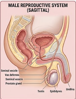
Figure: Gross anatomy of the male reproductive system showing testes,
ducts and accessory glands.
Microscopic Principle of Erection
Erection is primarily a vascular event. Parasympathetic stimulation promotes
relaxation of smooth muscle in erectile tissue and increases arterial inflow.
Blood fills the vascular spaces of erectile tissue, while venous outflow is
partly compressed, maintaining rigidity.
13. Functions of the Male Reproductive System
Function
Organ/Structure Mainly Involved
Description
Sperm production
Seminiferous tubules
Production of male gametes through spermatogenesis.
Sperm maturation
Epididymis
Sperm acquire functional maturity and motility.
Sperm transport
Vas deferens and ducts
Moves sperm toward the urethra.
Seminal fluid production
Seminal vesicles, prostate and bulbourethral glands
Provides fluid, nutrients and supportive substances.
Hormone secretion
Leydig and Sertoli cells
Testosterone and inhibin B contribute to reproductive regulation.
Copulation
Penis
Deposits semen within the female reproductive tract.
14. Female Reproductive System
The female reproductive system consists of the ovaries, uterine tubes,
uterus, cervix, vagina and external genitalia. The ovaries are the primary
female gonads. They produce oocytes and secrete reproductive hormones,
particularly estrogen and progesterone.
The uterine tubes receive the ovulated oocyte and provide the usual site of
fertilization. The fertilized ovum then travels toward the uterus, where
implantation occurs. The uterus supports development of the embryo and fetus
during pregnancy.
The cervix forms the lower portion of the uterus and connects it with the
vagina. The vagina serves as the copulatory canal and forms part of the
birth canal.
Figure: Gross anatomy of the female reproductive system.
15. Ovary
Location and Gross Anatomy
The ovaries are paired female gonads located in the pelvic cavity. They are
oval structures situated on either side of the uterus. Each ovary is attached
to surrounding structures by supporting ligaments and mesovarian tissue.
The ovary has two major anatomical regions: an outer
cortex and an inner medulla. The cortex
contains ovarian follicles at different stages of development, whereas the
medulla contains connective tissue, blood vessels, lymphatics and nerves.
Microscopic Anatomy
The ovarian cortex contains follicles consisting of an oocyte surrounded by
follicular cells. Follicles develop through primordial, primary, secondary
and mature stages. Following ovulation, the remaining follicular tissue
forms the corpus luteum.
The medulla consists largely of loose connective tissue and contains the
major vascular and neural structures supplying the ovary.
Figure: Gross and microscopic organization of the ovary showing cortex,
medulla and ovarian follicles.
Functions of Ovary
Production and maturation of female gametes.
Ovulation of a mature oocyte.
Secretion of estrogen.
Secretion of progesterone mainly from the corpus luteum after ovulation.
Secretion of inhibin.
Secretion of anti-Müllerian hormone (AMH) by granulosa cells.
Regulation of the menstrual and reproductive cycles.
16. Ovarian Follicles
An ovarian follicle consists of an oocyte and the surrounding supporting
follicular cells. Follicular development is a continuous process regulated
by gonadotropins and ovarian hormones.
Follicle
Important Characteristics
Primordial follicle
Primary oocyte surrounded by a single layer of flattened follicular cells.
Primary follicle
Follicular cells become cuboidal and proliferate; zona pellucida develops.
Secondary/antral follicle
Fluid-filled antrum develops and granulosa/theca layers become prominent.
Mature/Graafian follicle
Large follicle containing the mature oocyte and prominent antrum.
Corpus luteum
Post-ovulatory structure that secretes mainly progesterone and also estrogen.
Corpus albicans
Fibrous remnant formed when the corpus luteum regresses.
Granulosa and Theca Cells
Granulosa cells surround the developing oocyte and are important in follicular
support and estrogen production. Theca cells surrounding the follicle provide
androgen precursors that granulosa cells can convert into estrogens under
the influence of FSH.
17. Uterine Tube (Fallopian Tube)
The uterine tubes are paired muscular tubes extending from the region of the
ovaries toward the uterus. They provide the route through which the oocyte
travels toward the uterine cavity.
Parts of Uterine Tube
Part
Description
Infundibulum
Funnel-shaped lateral end with finger-like fimbriae.
Ampulla
Widest part and usual site of fertilization.
Isthmus
Narrow muscular segment close to the uterus.
Uterine/intramural part
Passes through the wall of the uterus.
Microscopic Anatomy
The mucosa is highly folded and lined mainly by simple columnar epithelium.
Two important epithelial cell types are ciliated cells and secretory cells.
Ciliary movement helps transport the oocyte toward the uterus, while secretory
cells contribute fluid that supports gamete and early embryo survival.
The muscular wall produces contractions that also assist movement of the
oocyte and early embryo.
Functions
Receives the ovulated oocyte.
Provides the usual site of fertilization.
Provides an environment for early embryo development.
Transports the embryo toward the uterus.
Clinical correlation:
Implantation outside the normal uterine cavity is called
ectopic pregnancy. The uterine tube is a common site of
ectopic implantation.
18. Uterus
The uterus is a hollow, muscular, pear-shaped organ located in the pelvis
between the urinary bladder and rectum. It is the organ in which implantation
normally occurs and where the embryo and fetus develop during pregnancy.
Gross Parts of Uterus
Part
Description
Fundus
Dome-shaped upper portion above the uterine tube openings.
Body
Main central portion of uterus.
Isthmus
Narrow region between body and cervix.
Cervix
Lower cylindrical portion projecting into the vagina.
Layers of Uterine Wall
The uterine wall consists of three major layers:
perimetrium, myometrium and endometrium.
1. Perimetrium
The perimetrium is the outer serosal covering of much of the uterus. It is
continuous with the surrounding peritoneal covering where applicable.
2. Myometrium
The myometrium is the thick middle layer composed predominantly of smooth
muscle. It enlarges substantially during pregnancy and generates powerful
contractions during labor.
3. Endometrium
The endometrium is the inner mucosal lining of the uterus. It undergoes
cyclic changes in response to ovarian hormones and provides the site for
implantation of the developing embryo.
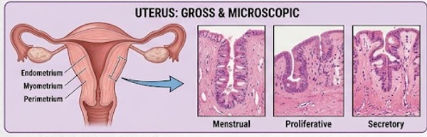
Figure: Gross structure of uterus and microscopic organization of the
uterine wall.
Functions of Uterus
Receives the implanted embryo.
Supports placental development.
Provides an environment for fetal development.
Contributes to menstruation when pregnancy does not occur.
Produces muscular contractions during childbirth.
19. Endometrium and Menstrual Cycle
The endometrium consists of a superficial functional layer and a deeper
basal layer. The functional layer undergoes cyclic changes and is shed
during menstruation when pregnancy does not occur. The basal layer remains
and contributes to regeneration of the endometrium.
Major Phases of Menstrual Cycle
Phase
Main Hormonal Influence
Main Endometrial Change
Menstrual phase
Withdrawal of ovarian hormones
Shedding of functional endometrium.
Proliferative phase
Estrogen
Regeneration and proliferation of endometrium.
Secretory phase
Progesterone
Endometrial glands become active and secretory.
If fertilization and implantation occur, hormonal support maintains the
endometrium. If pregnancy does not occur, the corpus luteum degenerates,
progesterone and estrogen levels fall, and the functional endometrium is
shed as menstruation.
20. Cervix
The cervix is the lower part of the uterus and projects into the upper
vagina. It contains a cervical canal that connects the uterine cavity with
the vagina.
Microscopic Anatomy
The endocervical canal is lined mainly by mucus-secreting simple columnar
epithelium, whereas the ectocervix is lined by stratified squamous
epithelium. The junction between these epithelial regions is clinically
important and is known as the transformation zone.
Functions
Provides passage between uterus and vagina.
Produces cervical mucus.
Changes mucus properties during the menstrual cycle.
Provides a barrier against ascending infection.
Dilates during labor to permit passage of the fetus.
Clinical correlation:
The transformation zone of the cervix is an important site for development
of cervical epithelial abnormalities. Cervical screening is designed to
detect precancerous changes before invasive disease develops.
21. Vagina
The vagina is a fibromuscular canal extending from the cervix to the
external genitalia. It functions as the female copulatory canal and forms
part of the birth canal.
Microscopic Anatomy
The vaginal mucosa is lined by non-keratinized stratified squamous epithelium.
The underlying connective tissue contains blood vessels and elastic fibers.
The muscular layer consists mainly of smooth muscle arranged in different
orientations.
The vaginal epithelium contains glycogen-rich cells. Vaginal microbial
communities metabolize available substrates and contribute to an acidic
environment that helps limit growth of some pathogenic organisms.
Functions
Receives the penis during sexual intercourse.
Provides a passage for menstrual discharge.
Forms part of the birth canal.
Provides a protective barrier against microorganisms.
22. External Female Genitalia
The external female genital structures are collectively called the
vulva. They protect the openings of the reproductive and
urinary tracts and contribute to sexual function.
Structure
Function
Mons pubis
Fat-containing tissue over the pubic region that provides protection.
Labia majora
Outer skin folds providing protection to deeper structures.
Labia minora
Inner folds surrounding the vestibule.
Clitoris
Erectile sensory organ involved in sexual arousal.
Vestibule
Area containing the external openings of the urethra and vagina.
Greater vestibular glands
Produce mucus that contributes to lubrication.
23. Mammary Glands
The mammary glands are specialized glands of the anterior thoracic wall.
Although they are not reproductive organs in the strict anatomical sense,
they are functionally associated with reproduction because they produce milk
after childbirth.
Gross Organization
Each mammary gland contains glandular tissue organized into lobes and
lobules. Milk produced by secretory alveoli passes through ducts toward
the nipple.
Microscopic Structure
The secretory units consist of alveoli lined by milk-secreting epithelial
cells and surrounded by myoepithelial cells. Myoepithelial contraction helps
expel milk into the duct system.
Hormonal Control
Hormone
Main Role
Estrogen
Promotes ductal development.
Progesterone
Promotes development of lobuloalveolar structures.
Prolactin
Stimulates milk synthesis.
Oxytocin
Causes contraction of myoepithelial cells and milk ejection.
24. Functions of Female Reproductive System
Function
Main Structure
Description
Oocyte production
Ovary
Produces and supports maturation of female gametes.
Hormone secretion
Ovary
Produces estrogen, progesterone, inhibin and AMH.
Fertilization
Uterine tube
Usually occurs in the ampulla.
Implantation
Uterus
Embryo implants into the endometrium.
Pregnancy
Uterus
Supports fetal growth and placental function.
Childbirth
Uterus and vagina
Uterine contractions and birth canal permit delivery.
Lactation
Mammary glands
Produces and delivers milk to the infant.
25. Hormonal Regulation of Reproductive System
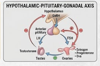
Reproductive function is regulated by an integrated endocrine pathway called
the hypothalamic-pituitary-gonadal axis. The hypothalamus
releases gonadotropin-releasing hormone (GnRH) in a pulsatile manner.
GnRH stimulates the anterior pituitary gland to release follicle-stimulating
hormone (FSH) and luteinizing hormone (LH).
In males, LH stimulates Leydig cells to produce testosterone, while FSH acts
mainly on Sertoli cells and supports spermatogenesis. Testosterone and
inhibin B provide feedback to the hypothalamus and pituitary.
In females, FSH promotes follicular development, while LH acts on theca cells,
supports ovulation and contributes to corpus luteum formation. Estrogen,
progesterone and inhibin participate in feedback regulation of the axis.
Figure: Hypothalamic-pituitary-gonadal axis regulating male and female
reproductive functions.
Male Hormonal Regulation
Hormone
Target
Major Function
GnRH
Anterior pituitary
Stimulates FSH and LH secretion.
LH
Leydig cells
Stimulates testosterone production.
FSH
Sertoli cells
Supports spermatogenesis and Sertoli cell function.
Testosterone
Male reproductive tissues and other target tissues
Supports male differentiation, reproductive function and secondary sexual characteristics.
Inhibin B
Anterior pituitary
Provides negative feedback mainly on FSH secretion.
Female Hormonal Regulation
Hormone
Main Target
Major Function
FSH
Ovarian follicles
Promotes follicular growth and granulosa cell function.
LH
Theca cells/corpus luteum
Supports androgen production, ovulation and luteal function.
Estrogen
Female reproductive tissues
Supports reproductive tract development and endometrial proliferation.
Progesterone
Uterus and other reproductive tissues
Supports secretory endometrium and pregnancy.
Inhibin
Anterior pituitary
Suppresses FSH secretion.
26. Gametogenesis
Spermatogenesis
Spermatogenesis is the process by which diploid spermatogenic cells develop
into haploid spermatozoa within the seminiferous tubules. It begins at
puberty and normally continues throughout adult life, although the rate
may decline with age.
Oogenesis
Oogenesis is the development of female gametes in the ovary. Unlike
spermatogenesis, the formation of primary oocytes begins before birth.
During reproductive life, selected follicles resume development during
each menstrual cycle. Usually one dominant follicle releases an oocyte
during ovulation.
Feature
Spermatogenesis
Oogenesis
Site
Seminiferous tubules
Ovarian follicles
Begins
At puberty
Before birth; cyclic maturation begins after puberty
Gamete produced
Spermatozoon
Oocyte
Number from one primary cell
Four haploid sperm cells
One functional ovum/oocyte plus polar bodies
Production pattern
Continuous after puberty
Cyclic and limited by ovarian reserve
27. Fertilization and Implantation
Fertilization is the process by which a spermatozoon fuses with the female
gamete to form a diploid zygote. It usually occurs in the ampulla of the
uterine tube.
Major Events of Fertilization
Sperm transport through the female reproductive tract.
Capacitation of sperm within the female reproductive tract.
Interaction of sperm with the oocyte coverings.
Acrosomal reaction and penetration of the zona pellucida.
Fusion of sperm and oocyte membranes.
Prevention of polyspermy.
Completion of oocyte meiosis II.
Fusion of parental genetic material to establish the zygote.
The developing embryo undergoes cleavage while moving toward the uterus.
After reaching the blastocyst stage, it attaches to and implants within
the endometrium.
Exam point:
The usual site of fertilization is the ampulla of the uterine tube,
whereas the normal site of implantation is the
endometrium of the uterus.
28. Important Reproductive Hormones
Hormone
Major Source
Main Function
Testosterone
Leydig cells of testis
Male reproductive development, secondary sexual characteristics and support of spermatogenesis.
Estrogen
Ovarian follicles; placenta during pregnancy
Female reproductive development, endometrial proliferation and bone maintenance.
Progesterone
Corpus luteum; placenta during pregnancy
Supports secretory endometrium and pregnancy.
Inhibin
Sertoli cells and ovarian granulosa cells
Suppresses FSH secretion.
AMH
Granulosa cells; Sertoli cells in fetal male
Important in follicular physiology and fetal reproductive tract development.
hCG
Placental trophoblast
Maintains corpus luteum early in pregnancy.
Prolactin
Anterior pituitary
Stimulates milk synthesis.
Oxytocin
Posterior pituitary release
Promotes uterine contraction and milk ejection.
29. Clinical Correlations
1. Male Infertility
Male infertility may result from abnormalities in sperm production, sperm
motility, sperm morphology, ductal obstruction, endocrine dysfunction or
other reproductive conditions. Evaluation commonly includes semen analysis
and assessment of reproductive history.
2. Benign Prostatic Hyperplasia
Benign prostatic hyperplasia is non-malignant enlargement of the prostate
that commonly occurs with aging. Because the prostate surrounds the proximal
urethra, enlargement can cause lower urinary tract symptoms such as weak
stream, hesitancy and incomplete emptying.
3. Cryptorchidism
Cryptorchidism refers to failure of one or both testes to descend into the
scrotum. Increased temperature in an undescended testis can impair normal
spermatogenesis and is associated with increased risk of certain testicular
tumors.
4. Polycystic Ovary Syndrome
Polycystic ovary syndrome is a common endocrine and reproductive disorder
associated with ovulatory dysfunction and variable androgen excess. It may
present with irregular menstrual cycles, clinical or biochemical
hyperandrogenism and difficulty with fertility. The term should not be
interpreted as simply meaning multiple ovarian cysts.
5. Endometriosis
Endometriosis is a condition in which tissue resembling endometrial tissue
is present outside the uterine cavity. It may cause pelvic pain, painful
menstruation and infertility.
6. Ectopic Pregnancy
An ectopic pregnancy occurs when implantation takes place outside the normal
uterine cavity. The uterine tube is a common location. Rupture of a tubal
ectopic pregnancy can cause severe internal bleeding and is a medical
emergency.
7. Cervical Cancer
Cervical cancer commonly develops from persistent infection with
high-risk human papillomavirus (HPV) types followed by progressive cellular
changes. The transformation zone is particularly important in cervical
screening.
8. Erectile Dysfunction
Erectile dysfunction is the persistent inability to achieve or maintain an
erection sufficient for satisfactory sexual activity. It may have vascular,
neurological, endocrine, medication-related or psychological contributors.
30. 10-Mark Long Questions
Long Question 1: Describe the male reproductive system with its gross anatomy, microscopic anatomy and functions.
Introduction:
The male reproductive system is composed of paired testes, ducts, accessory
reproductive glands and external genitalia. Its major functions are production
of sperm, secretion of male reproductive hormones and delivery of sperm into
the female reproductive tract.
1. Testes:
The testes are paired male gonads located in the scrotum. Each testis is
covered by tunica vaginalis and tunica albuginea. Septa from the tunica
albuginea divide the testis into lobules containing seminiferous tubules.
The seminiferous tubules are lined by germ cells and Sertoli cells. Between
the tubules are Leydig cells that produce testosterone.
2. Epididymis:
The epididymis is a highly coiled duct divided into head, body and tail.
It is lined by pseudostratified columnar epithelium with stereocilia.
It is the major site for sperm maturation, acquisition of motility and
storage.
3. Vas deferens:
The vas deferens is a thick muscular duct that transports sperm from the
epididymis to the ejaculatory duct. Its strong muscular wall produces
peristaltic contractions during ejaculation.
4. Accessory glands:
The seminal vesicles produce fructose-rich secretion. The prostate produces
prostatic fluid containing substances that support semen function. The
bulbourethral glands produce mucus-rich fluid that lubricates the urethra.
5. Penis:
The penis contains erectile tissues, mainly corpora cavernosa and corpus
spongiosum. It acts as the male copulatory organ and provides a passage for
semen.
Functions:
The male reproductive system produces spermatozoa, secretes testosterone,
provides sperm maturation and transport, produces seminal fluid and permits
sexual intercourse and deposition of semen.
Long Question 2: Describe the microscopic structure of testis and explain spermatogenesis.
Introduction:
The testis performs both exocrine and endocrine functions. Its exocrine
function is sperm production, while its endocrine function is mainly
testosterone secretion.
Microscopic structure:
The testis is divided into lobules by connective tissue septa. Each lobule
contains seminiferous tubules. The seminiferous epithelium consists of
developing germ cells supported by Sertoli cells. Leydig cells occur in the
interstitial tissue between seminiferous tubules.
Spermatogenesis:
Spermatogenesis begins with spermatogonia. These cells develop into primary
spermatocytes, which undergo meiosis I to form secondary spermatocytes.
Secondary spermatocytes undergo meiosis II and form haploid spermatids.
Spermatids then undergo spermiogenesis and become spermatozoa.
Sertoli cell functions:
Sertoli cells support germ cells, contribute to the blood-testis barrier,
provide nutrients to developing sperm, participate in spermiogenesis and
secrete inhibin B and androgen-binding protein.
Leydig cell function:
Leydig cells produce testosterone in response to LH. Testosterone supports
male reproductive development, secondary sexual characteristics and
spermatogenesis.
Long Question 3: Describe the female reproductive system with its gross and microscopic anatomy and functions.
Introduction:
The female reproductive system consists of ovaries, uterine tubes, uterus,
cervix, vagina and external genitalia. It produces female gametes and
hormones and provides the anatomical environment for fertilization,
implantation, pregnancy and childbirth.
Ovaries:
The ovaries are paired female gonads. Their cortex contains ovarian follicles
at different stages of development, while the medulla contains connective
tissue, blood vessels and nerves. Ovaries produce oocytes and secrete estrogen,
progesterone, inhibin and AMH.
Uterine tubes:
The uterine tubes consist of infundibulum, ampulla, isthmus and intramural
parts. The ampulla is the usual site of fertilization. The mucosa contains
ciliated and secretory cells that support gamete and embryo transport.
Uterus:
The uterus is divided into fundus, body, isthmus and cervix. Its wall consists
of perimetrium, myometrium and endometrium. The endometrium undergoes cyclic
changes and provides the site for implantation.
Vagina:
The vagina is a fibromuscular canal lined by non-keratinized stratified
squamous epithelium. It acts as the copulatory canal, passage for menstrual
flow and part of the birth canal.
Functions:
The female reproductive system produces oocytes, secretes reproductive
hormones, receives sperm, provides the site of fertilization, supports
implantation and fetal development and participates in childbirth.
Long Question 4: Describe the ovary with its gross and microscopic structure and explain its functions.
Gross anatomy:
The ovaries are paired oval organs situated in the pelvic cavity on either
side of the uterus. They are attached to surrounding structures by supporting
ligaments and mesovarian tissue.
Microscopic anatomy:
The ovary has an outer cortex and inner medulla. The cortex contains follicles
at different developmental stages. The medulla contains loose connective
tissue, vessels, lymphatics and nerves.
Follicular development:
Follicles progress from primordial to primary, secondary and mature stages.
Following ovulation, the remaining follicular tissue forms the corpus luteum,
which secretes progesterone and estrogen. If pregnancy does not occur, it
regresses and eventually forms a corpus albicans.
Functions:
The ovary produces and supports maturation of female gametes and secretes
estrogen, progesterone, inhibin and AMH. These hormones regulate the
menstrual cycle, reproductive tract function and development of female
secondary sexual characteristics.
Long Question 5: Describe the uterus with its gross and microscopic anatomy and functions.
Gross structure:
The uterus is a hollow muscular organ divided into fundus, body, isthmus and
cervix. It lies between the urinary bladder and rectum.
Uterine wall:
The uterine wall consists of perimetrium, myometrium and endometrium.
The perimetrium forms the outer covering, the myometrium is composed of
smooth muscle, and the endometrium forms the inner mucosa.
Endometrium:
The endometrium undergoes cyclic changes under the influence of ovarian
hormones. During the proliferative phase, estrogen promotes regeneration and
growth. During the secretory phase, progesterone prepares the endometrium
for possible implantation.
Functions:
The uterus receives the implanted embryo, supports placental and fetal
development, undergoes cyclic menstrual changes and produces contractions
during labor.
Long Question 6: Compare the male and female reproductive systems.
Feature
Male
Female
Primary gonad
Testis
Ovary
Gamete
Sperm
Oocyte
Main sex hormone
Testosterone
Estrogen and progesterone
Gamete production
Begins at puberty and is relatively continuous
Begins before birth; cyclic maturation after puberty
Accessory glands
Seminal vesicles, prostate, bulbourethral glands
No direct equivalent group of seminal glands
Fertilization
Produces and delivers sperm
Usually occurs in ampulla of uterine tube
Pregnancy
Does not provide fetal environment
Uterus supports pregnancy
Childbirth
Not directly involved
Uterus and vagina participate
Long Question 7: Explain hormonal regulation of the reproductive system.
Hypothalamus:
The hypothalamus releases GnRH in pulses. GnRH acts on the anterior pituitary
to stimulate secretion of FSH and LH.
Male:
LH stimulates Leydig cells to produce testosterone. FSH acts on Sertoli cells
and supports spermatogenesis. Testosterone and inhibin B provide negative
feedback to regulate the axis.
Female:
FSH stimulates follicular development and granulosa cell activity. LH
supports theca cell androgen production, triggers ovulation and supports
corpus luteum function. Estrogen, progesterone and inhibin regulate the
hypothalamic-pituitary system through feedback.
Importance:
The HPG axis coordinates gonadal development, gamete production, reproductive
cycles, sexual development and reproductive hormone secretion.
Describe their location and functions in spermatogenesis and endocrine regulation.
3. Leydig Cells
Location, hormonal regulation and functions of testosterone secretion.
4. Epididymis
Gross structure, histology and role in sperm maturation.
5. Seminal Vesicle
Structure and composition/functions of seminal secretion.
6. Prostate Gland
Gross anatomy, histology, secretion and clinical significance.
7. Ovary
Gross structure, cortex, medulla, follicles and endocrine functions.
8. Ovarian Follicles
Explain follicular development from primordial follicle to corpus luteum.
9. Uterine Tube
Parts, microscopic structure and functions.
10. Uterus
Parts and three layers of uterine wall with functions.
11. Endometrium
Functional and basal layers and cyclic changes.
12. Cervix
Gross and microscopic anatomy and functions.
13. Vagina
Histology and major functions.
14. Spermatogenesis
Explain stages from spermatogonium to spermatozoon.
15. Oogenesis
Explain formation and maturation of female gamete.
16. HPG Axis
Explain GnRH, FSH, LH and gonadal hormones.
32. Important 2-Mark Questions
Question
Key Answer
Define reproductive system.
Group of organs responsible for gamete production, reproductive hormone secretion and reproduction.
Name the male gonad.
Testis.
Name the female gonad.
Ovary.
What is the function of testis?
Sperm production and secretion of male reproductive hormones, mainly testosterone.
What are seminiferous tubules?
Highly coiled tubules in the testis where spermatogenesis occurs.
What are Sertoli cells?
Supporting cells of seminiferous tubules involved in germ-cell support and reproductive regulation.
What are Leydig cells?
Interstitial endocrine cells that secrete testosterone.
Function of epididymis?
Sperm maturation, storage and transport.
Name three male accessory glands.
Seminal vesicles, prostate and bulbourethral glands.
Function of seminal vesicle?
Produces fructose-rich seminal fluid that contributes to semen.
Function of prostate?
Produces prostatic fluid that contributes to semen and supports sperm function.
Name the female reproductive organs.
Ovaries, uterine tubes, uterus, cervix, vagina and external genitalia.
What is the function of ovary?
Produces oocytes and secretes ovarian hormones.
What is the usual site of fertilization?
Ampulla of the uterine tube.
What is the site of implantation?
Endometrium of the uterus.
Name the layers of uterine wall.
Perimetrium, myometrium and endometrium.
What is myometrium?
Thick smooth muscle layer of the uterus.
What is endometrium?
Inner mucosal lining of the uterus involved in cyclic changes and implantation.
What is corpus luteum?
Post-ovulatory ovarian structure that mainly secretes progesterone.
Name two major ovarian hormones.
Estrogen and progesterone.
What is testosterone?
Major male sex steroid hormone produced mainly by Leydig cells.
What is estrogen?
Major group of female sex steroid hormones involved in reproductive development and function.
What is progesterone?
Ovarian steroid hormone important for secretory endometrium and pregnancy support.
Define spermatogenesis.
Process by which spermatogenic cells develop into mature spermatozoa.
Define oogenesis.
Process of development and maturation of female gametes in the ovary.
What is GnRH?
Hypothalamic hormone that stimulates pituitary secretion of FSH and LH.
Function of FSH in male?
Acts mainly on Sertoli cells and supports spermatogenesis.
Function of LH in male?
Stimulates Leydig cells to produce testosterone.
Function of FSH in female?
Promotes ovarian follicular development.
Function of LH in female?
Participates in ovulation and corpus luteum formation.
33. High-Yield Examination Tables
Male Reproductive Organs at a Glance
Organ
Key Structure
Main Function
Testis
Seminiferous tubules
Sperm production and testosterone secretion.
Epididymis
Coiled duct with stereocilia
Sperm maturation and storage.
Vas deferens
Thick muscular wall
Sperm transport.
Seminal vesicle
Highly folded gland
Fructose-rich seminal secretion.
Prostate
Tubuloalveolar glands
Prostatic secretion.
Bulbourethral gland
Mucus-secreting gland
Lubrication and urethral preparation.
Penis
Erectile tissue
Copulation and semen delivery.
Female Reproductive Organs at a Glance
Organ
Key Structure
Main Function
Ovary
Follicles and corpus luteum
Oocyte development and hormone secretion.
Uterine tube
Ciliated mucosa
Oocyte/embryo transport and usual site of fertilization.
Uterus
Endometrium and myometrium
Implantation and fetal development.
Cervix
Cervical canal
Mucus production, barrier and birth canal contribution.
Vagina
Stratified squamous epithelium
Copulation, menstrual passage and birth canal.
Mammary gland
Alveoli and ducts
Milk production and delivery.
Male vs Female Gametogenesis
Point
Spermatogenesis
Oogenesis
Site
Testis
Ovary
Starting time
Puberty
Fetal life; maturation resumes after puberty
Pattern
Relatively continuous
Cyclic
Functional gametes
Four sperm from one primary spermatocyte
One functional gamete from one primary oocyte
Motility
Sperm are motile
Oocyte is non-motile and transported by tube mechanisms
Reproductive Hormones
Hormone
Source
Major Function
GnRH
Hypothalamus
Stimulates FSH and LH secretion.
FSH
Anterior pituitary
Spermatogenesis support and ovarian follicular development.
LH
Anterior pituitary
Testosterone production, ovulation and luteal function.
Testosterone
Leydig cells
Male reproductive development and function.
Estrogen
Ovarian follicles
Female reproductive development and endometrial proliferation.
Progesterone
Corpus luteum
Secretory endometrium and pregnancy support.
Inhibin
Gonads
Negative feedback mainly on FSH.
hCG
Placenta
Maintains corpus luteum during early pregnancy.
Prolactin
Anterior pituitary
Milk synthesis.
Oxytocin
Posterior pituitary release
Uterine contraction and milk ejection.
34. Important Diagrams to Practice
For MSc/TU examinations, diagrams should be simple, clearly labeled and
directly related to the answer. The following diagrams are especially useful
for long questions.
Diagram 1
Complete male reproductive system.
Diagram 2
Gross and microscopic structure of testis.
Diagram 3
Seminiferous tubule and spermatogenesis.
Diagram 4
Structure of spermatozoon.
Diagram 5
Complete female reproductive system.
Diagram 6
Gross and microscopic structure of ovary.
Diagram 7
Stages of ovarian follicular development.
Diagram 8
Gross and microscopic structure of uterus.
Diagram 9
Hypothalamic-pituitary-gonadal axis.
Diagram 10
Menstrual cycle and hormonal changes.
35. How to Write a Reproductive System Long Answer
Step 1 — Definition:
Start with a clear definition of the reproductive system.
Step 2 — Classification:
Mention male and female reproductive systems and identify the primary gonads.
Step 3 — Gross anatomy:
Describe location, major parts and important anatomical relationships.
Step 4 — Microscopic anatomy:
Mention the important tissue layers, epithelial lining, specialized cells,
follicles or seminiferous tubules.
Step 5 — Functions:
Explain the function of each major organ rather than simply listing names.
Step 6 — Hormonal role:
For gonads, mention the important reproductive hormones and their major roles.
Step 8 — Clinical correlation:
If space and time permit, add one or two relevant clinical conditions.
High-scoring approach:
For a 10-mark question, use headings and subheadings. A long answer should
look organized rather than being written as one continuous paragraph.
Include both gross and microscopic anatomy because both are specifically
included in the MB 551 syllabus.
36. Last-Minute Revision
Testes are the male gonads.
Ovaries are the female gonads.
Seminiferous tubules are the site of spermatogenesis.
Sertoli cells support developing germ cells.
Leydig cells secrete testosterone.
Epididymis is the major site of sperm maturation and storage.
Vas deferens transports sperm.
Seminal vesicles produce fructose-rich seminal fluid.
Prostate contributes prostatic fluid to semen.
Bulbourethral glands produce mucus-rich secretion.
The penis is the male copulatory organ.
Ovarian cortex contains developing follicles.
Ovarian medulla contains vessels, nerves and connective tissue.
Granulosa and theca cells participate in ovarian hormone production.
Ampulla of uterine tube is the usual site of fertilization.
Uterus is the usual site of implantation.
Uterine wall consists of perimetrium, myometrium and endometrium.
Myometrium produces uterine contractions.
Endometrium undergoes cyclic changes.
Cervix connects uterus and vagina.
Vagina is lined mainly by non-keratinized stratified squamous epithelium.
GnRH stimulates pituitary secretion of FSH and LH.
FSH supports follicular development and Sertoli-cell function.
LH stimulates Leydig cells and participates in ovulation/luteal function.
Testosterone is the major male sex steroid.
Estrogen and progesterone are major ovarian steroid hormones.
Progesterone supports the secretory endometrium and pregnancy.
Inhibin suppresses FSH secretion.
hCG maintains the corpus luteum during early pregnancy.
Prolactin stimulates milk synthesis.
Oxytocin promotes uterine contraction and milk ejection.
Spermatogenesis is relatively continuous after puberty.
Oocyte maturation is cyclic during reproductive life.
Ectopic pregnancy commonly involves the uterine tube.
BPH can obstruct the male urethra.
Cervical screening focuses on detecting abnormal changes in cervical epithelium.
37. Chapter Summary
The reproductive system is composed of male and female reproductive organs
that collectively perform gamete production, reproductive hormone secretion,
fertilization and, in females, pregnancy and childbirth. The male reproductive
system consists mainly of the testes, ducts, accessory glands and penis.
The testes contain seminiferous tubules for spermatogenesis and Leydig cells
for testosterone secretion.
The female reproductive system consists mainly of ovaries, uterine tubes,
uterus, cervix, vagina and external genitalia. The ovaries contain follicles
and function as both gamete-producing and endocrine organs. The uterine tubes
transport the oocyte and are the usual site of fertilization. The uterus
provides the site for implantation and fetal development, while the vagina
functions as the copulatory canal and part of the birth canal.
Both reproductive systems are regulated by the hypothalamic-pituitary-gonadal
axis. GnRH from the hypothalamus regulates pituitary FSH and LH secretion,
which in turn control gonadal activity. Testosterone, estrogen, progesterone
and inhibin provide feedback regulation. Understanding the relationship
between gross anatomy, microscopic anatomy, hormonal regulation and
function is essential for answering MB 551 long questions effectively.