UNIT 7 — SPECTROSCOPY
7.1 Introduction to Spectroscopy
Spectroscopy is one of the most important analytical approaches in biochemistry, microbiology, molecular biology, biotechnology, medicine and pharmaceutical science. Biological molecules interact with electromagnetic radiation in characteristic ways. These interactions can be measured and used to obtain information about concentration, structure, chemical environment and molecular interactions.
Different regions of the electromagnetic spectrum provide different types of information. Ultraviolet and visible spectroscopy is frequently used for quantitative measurement of biological compounds, while infrared spectroscopy provides information about molecular vibrations. Fluorescence spectroscopy is highly sensitive and is useful for studying fluorescent molecules and fluorescently labelled biological systems.
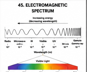
7.2 Electromagnetic Radiation
Electromagnetic radiation has both wave-like and particle-like properties. It can be described in terms of wavelength, frequency and energy.
where:
- c = speed of light
- λ = wavelength
- ν = frequency
The energy of electromagnetic radiation can be expressed as:
where h is Planck's constant.
Therefore, higher-frequency radiation has higher energy, while longer wavelength radiation has lower energy.
7.3 Interaction of Radiation with Matter
When electromagnetic radiation encounters a substance, several things may occur. Depending on the wavelength and molecular structure, the radiation may be transmitted, absorbed, reflected, scattered or emitted.
7.4 Absorption Spectroscopy
In absorption spectroscopy, the sample absorbs radiation of particular wavelengths. The amount of radiation absorbed can be related to the concentration of the absorbing substance under appropriate conditions.
7.5 Beer-Lambert Law
One of the most important quantitative relationships in spectrophotometry is the Beer-Lambert law.
where:
- A = absorbance
- ε = molar absorptivity
- l = path length of cuvette
- c = concentration of absorbing substance
The law indicates that, within its range of validity, absorbance is proportional to concentration and path length.
7.6 UV-Visible Spectrophotometry
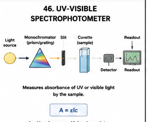
UV-visible spectrophotometry measures the absorption of ultraviolet and visible radiation by a sample. It is among the most widely used spectroscopic techniques in biological laboratories.
Principle
When a molecule absorbs UV or visible radiation, electrons may undergo transitions between different energy levels. The wavelength and intensity of absorption depend on the molecular structure and concentration.
Main Components
- Radiation source
- Monochromator or wavelength-selection system
- Sample holder or cuvette
- Detector
- Signal processing system
- Display or computer system
Sources
Different radiation sources may be used for different wavelength regions. UV measurements commonly require a source appropriate for ultraviolet radiation, while visible measurements require a suitable visible-light source.
Monochromator
The monochromator or wavelength-selection system isolates a relatively narrow range of wavelengths from the source. This allows the sample to be examined at a selected wavelength.
Cuvettes
The sample is placed in a cuvette. The material of the cuvette must be appropriate for the wavelength region being measured.
Detector
The detector converts the transmitted radiation into an electrical signal, which is processed to provide absorbance or related measurements.
7.7 General Working of a Spectrophotometer
7.8 Applications of UV-Visible Spectrophotometry
- Measurement of nucleic-acid concentration
- Protein concentration estimation using suitable assays
- Measurement of enzyme activity
- Monitoring biochemical reactions
- Determination of pigment concentration
- Growth estimation in microbial cultures by optical density
- Quantitative analysis of colored compounds
- Drug and pharmaceutical analysis
- Determination of reaction kinetics
7.9 Advantages
- Rapid analysis
- Simple operation for routine measurements
- Non-destructive in many applications
- Requires relatively small sample volumes
- Suitable for quantitative measurements
- Can be automated
7.10 Limitations
- Interfering substances can affect absorbance.
- Highly concentrated samples may fall outside the useful linear range.
- Turbidity can cause scattering and inaccurate results.
- Incorrect blanking can introduce systematic error.
- The selected wavelength must be appropriate for the analyte.
7.11 Spectrophotometric Numerical Problems
Spectroscopy may produce numerical questions based on the Beer-Lambert law. Students should be able to rearrange the equation and calculate absorbance, concentration, path length or molar absorptivity when the other quantities are provided.
Example 1 — Finding Absorbance
If:
- ε = 10,000 L mol⁻¹ cm⁻¹
- l = 1 cm
- c = 2 × 10⁻⁵ mol/L
Example 2 — Finding Concentration
Suppose:
- Absorbance = 0.60
- ε = 12,000 L mol⁻¹ cm⁻¹
- l = 1 cm
Example 3 — Finding Path Length
If A = 0.80, ε = 16,000 L mol⁻¹ cm⁻¹ and c = 5 × 10⁻⁵ mol/L:
Calculation Tips
- Write Beer-Lambert law before substituting values.
- Check the units of ε, l and concentration.
- Convert concentration to the appropriate units.
- Rearrange the equation carefully.
- Always include the final unit where applicable.
- Check whether the calculated value is physically reasonable.
7.12 Fluorescence Spectroscopy
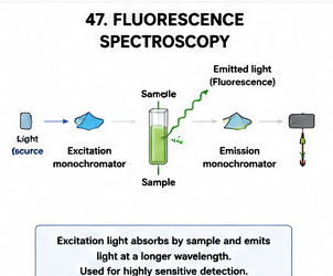
Fluorescence spectroscopy measures light emitted by a substance after it has absorbed radiation of an appropriate wavelength.
Principle
A fluorescent molecule absorbs energy and is promoted to an excited electronic state. It subsequently loses some energy and returns toward a lower energy state while emitting light. The emitted light generally has a longer wavelength than the excitation radiation.
Stokes Shift
The difference between the wavelength of excitation and the wavelength of maximum emission is commonly described as the Stokes shift.
Applications
- Fluorescent labelling
- Protein studies
- DNA and RNA detection
- Cell imaging
- Immunofluorescence
- Enzyme and molecular interaction studies
- Diagnostic assays
7.13 Infrared Spectroscopy
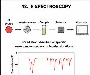
Infrared spectroscopy examines the interaction of infrared radiation with molecules. Absorption of infrared radiation is associated with molecular vibrational transitions.
Principle
Chemical bonds can undergo vibrational motions such as stretching and bending. When the frequency of incident infrared radiation corresponds to an allowed molecular vibration, absorption can occur.
Applications
- Identification of functional groups
- Characterization of organic compounds
- Biomolecular analysis
- Detection of chemical changes
- Quality control
- Research on proteins, lipids and carbohydrates
7.14 Comparison of Important Spectroscopic Techniques
| Technique | Major interaction | Major information | Important application |
|---|---|---|---|
| UV-Visible | Electronic absorption | Concentration and electronic transitions | Quantitative biochemical analysis |
| Fluorescence | Excitation and emission | Highly sensitive molecular detection | Labelling and molecular studies |
| Infrared | Molecular vibrations | Functional groups and chemical structure | Compound characterization |
Important Examination Questions — Unit 7
UNIT 8 — MOLECULAR TECHNIQUES
8.1 Introduction
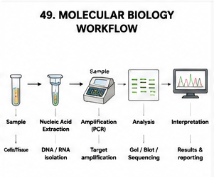
Molecular techniques are laboratory methods used to study nucleic acids, genes, genomes, gene expression and molecular interactions. These techniques have transformed microbiology, biotechnology, genetics, diagnostics and biomedical research.
Important molecular techniques include DNA extraction, RNA extraction, polymerase chain reaction, gel electrophoresis, restriction digestion, DNA amplification, cloning and nucleic-acid detection methods.
8.2 DNA Extraction
Basic Principle
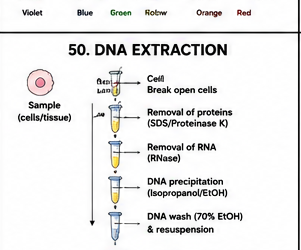
DNA extraction involves disruption of cells, release of nucleic acids, removal of proteins and other contaminants, and recovery of DNA in a suitable solution.
Major Steps
- Collection of biological material
- Cell disruption
- Cell lysis
- Release of DNA
- Removal of proteins and cellular contaminants
- DNA purification
- DNA recovery
- DNA quality assessment
Applications
- PCR
- DNA sequencing
- Genotyping
- Cloning
- Restriction analysis
- Microbial identification
- Forensic analysis
- Genetic research
8.3 RNA Extraction
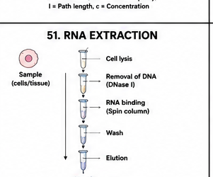
RNA extraction is used to isolate RNA from biological samples for applications such as gene-expression studies, reverse transcription and RNA sequencing.
Important Consideration
RNA is more susceptible to degradation than DNA. Therefore, careful handling and control of RNase contamination are essential during RNA isolation.
Applications
- Gene-expression analysis
- RT-PCR
- Quantitative PCR after reverse transcription
- RNA sequencing
- Transcriptomic studies
8.4 Polymerase Chain Reaction — PCR
Principle
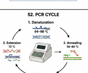
PCR uses a DNA template, primers, nucleotides, DNA polymerase, buffer and appropriate reaction conditions. Repeated cycles of heating and cooling allow the target sequence to be copied many times.
Major Components of PCR
- Template DNA
- Forward primer
- Reverse primer
- DNA polymerase
- dNTPs
- Reaction buffer
- Magnesium ions
- Water or appropriate reaction solvent
Three Fundamental PCR Steps
1. Denaturation
The double-stranded DNA template is separated into single strands by heating.
2. Annealing
The reaction is cooled to a temperature that allows primers to bind to their complementary target sequences.
3. Extension
DNA polymerase extends the primers by adding nucleotides to form new DNA strands.
8.5 PCR Amplification and Exponential Increase
Under idealized conditions, the amount of target DNA approximately doubles during each amplification cycle.
where:
- N = theoretical amount after n cycles
- N₀ = starting amount
- n = number of cycles
Example
If one starting molecule were amplified under ideal conditions for 10 cycles:
Thus, the idealized number of copies would be approximately 1024.
8.6 Applications of PCR
- Detection of microorganisms
- Identification of pathogens
- Genetic mutation analysis
- Cloning
- Forensic DNA analysis
- Research on gene structure
- Detection of specific DNA sequences
- Medical molecular diagnostics
- Food microbiology
- Environmental microbiology
8.7 Advantages of PCR
- Very sensitive
- Rapid compared with many traditional amplification methods
- Highly specific when primers are appropriately designed
- Requires relatively small amounts of starting material
- Can amplify specific target regions
8.8 Limitations of PCR
- Contamination can produce false-positive results.
- Poor primer design can reduce specificity.
- Inhibitory substances can interfere with amplification.
- PCR detects nucleic-acid targets and does not automatically establish viability of an organism.
- Optimization may be required for difficult targets.
8.9 Reverse Transcription PCR — RT-PCR
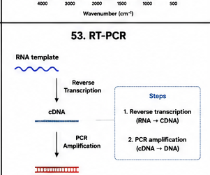
Reverse transcription PCR is used when the starting material is RNA. Because conventional DNA polymerases amplify DNA rather than RNA, RNA is first converted into complementary DNA (cDNA) using a reverse transcriptase enzyme.
Applications
- Gene-expression studies
- Detection of RNA viruses
- Analysis of messenger RNA
- Research on transcription
8.10 Quantitative PCR — qPCR
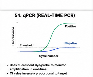
Quantitative PCR, often called real-time PCR, monitors amplification during the PCR process using fluorescence.
Basic Principle
Fluorescent signals increase as amplified DNA accumulates. The instrument records fluorescence during successive cycles, allowing amplification to be monitored in real time.
Applications
- Quantification of nucleic acids
- Gene-expression analysis
- Pathogen detection
- Clinical molecular diagnostics
- Research in gene regulation
8.11 Restriction Enzyme Analysis
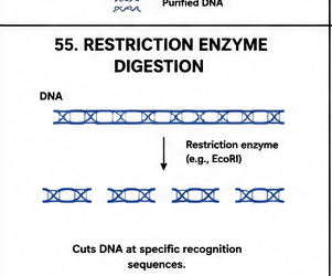
Restriction enzymes are nucleases that recognize particular DNA sequences and cleave DNA at or near specific recognition sites.
Applications
- DNA mapping
- Cloning
- Recombinant DNA technology
- DNA fragment analysis
- Genetic research
8.12 Sticky Ends and Blunt Ends
Depending on the cleavage pattern, restriction enzymes can produce overhanging single-stranded ends or blunt-ended DNA fragments.
| Feature | Sticky Ends | Blunt Ends |
|---|---|---|
| Structure | Single-stranded overhang | No overhanging bases |
| Base pairing | Can pair with complementary overhangs | No complementary overhang |
| Use in cloning | Often facilitates directional or compatible ligation strategies | Can also be ligated but may require different conditions |
8.13 DNA Cloning
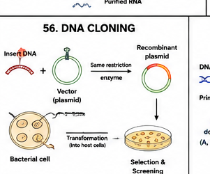
DNA cloning is the production of multiple copies of a DNA fragment by introducing the fragment into a suitable biological or molecular replication system.
General Steps
- Selection of target DNA
- Preparation of vector and insert
- Restriction digestion or other compatible DNA assembly strategy
- Joining of DNA fragments
- Introduction into host cells
- Selection of recombinant cells
- Screening and confirmation
- Propagation and analysis
Applications
- Gene analysis
- Protein production
- Recombinant DNA research
- Production of research constructs
- Study of gene function
8.14 DNA Sequencing
DNA sequencing determines the nucleotide order within a DNA molecule. Sequence information is fundamental for identifying genes, studying mutations and characterizing genomes.
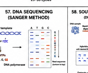
Applications
- Gene identification
- Mutation detection
- Microbial identification
- Genome analysis
- Evolutionary studies
- Clinical molecular investigations
- Research in genomics
8.15 Southern Blotting
Southern blotting is a molecular technique used to detect specific DNA sequences within a complex mixture of DNA fragments.

General Principle
DNA is separated by electrophoresis, transferred from the gel to a membrane and then detected using a complementary labelled probe.
Applications
- Detection of specific DNA sequences
- Gene organization studies
- Genetic analysis
- Research on gene rearrangements
8.16 Northern Blotting
Northern blotting is used to study specific RNA molecules. RNA is separated according to size, transferred to a membrane and detected using a complementary probe.
Applications
- Gene-expression studies
- RNA size analysis
- Detection of specific transcripts
- Studies of transcriptional regulation
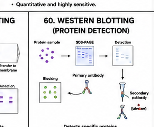
8.17 Western Blotting
Western blotting is used to detect a specific protein within a mixture of proteins. Proteins are separated by electrophoresis, transferred to a membrane and detected using antibodies.
General Steps
- Protein extraction
- Protein separation by PAGE
- Transfer of proteins to membrane
- Blocking
- Primary antibody incubation
- Secondary antibody incubation
- Detection
- Interpretation
Applications
- Protein detection
- Protein-expression studies
- Verification of recombinant proteins
- Research on disease-associated proteins
- Confirmation of protein identity
8.18 Comparison of Molecular Blotting Techniques
| Technique | Target | Detection principle |
|---|---|---|
| Southern blot | DNA | Hybridization with complementary probe |
| Northern blot | RNA | Hybridization with complementary probe |
| Western blot | Protein | Antibody-based detection |
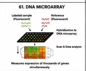
8.19 DNA Microarray
A DNA microarray contains many immobilized DNA probes arranged in an ordered pattern. It can be used to examine the presence or relative abundance of many nucleic-acid targets simultaneously.
Applications
- Gene-expression profiling
- Genotyping
- Comparative genomic studies
- Research on disease-associated genes
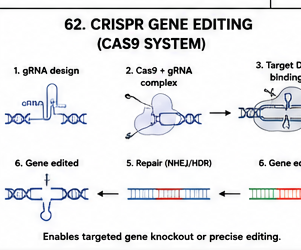
8.20 Basic Introduction to CRISPR-Based Molecular Technology
CRISPR-based systems are molecular tools derived from microbial defence mechanisms and adapted for targeted nucleic-acid manipulation. They are widely used in modern molecular biology research.
General Components
- Target DNA sequence
- Guide RNA
- CRISPR-associated nuclease
- Target recognition region
Applications
- Gene-function studies
- Genome engineering
- Research on genetic disorders
- Development of biological models
- Molecular biotechnology research
8.21 Nucleic-Acid Quality Assessment
Before PCR, sequencing, cloning or other molecular applications, the quality of DNA or RNA should be evaluated.
Important Parameters
- Concentration
- Purity
- Integrity
- Presence of inhibitors
- Suitability for downstream applications
8.22 DNA/RNA Quantification
Nucleic-acid concentration can be estimated by spectrophotometric methods or fluorescence-based methods depending on the sample and required sensitivity.
Purity Ratios
Absorbance ratios such as A260/A280 and A260/A230 can provide useful indications of nucleic-acid sample quality, although these values should be interpreted together with other evidence rather than treated as an absolute quality judgment.
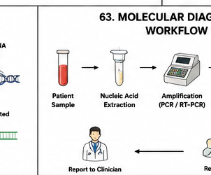
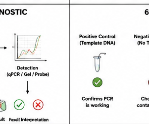
8.23 Molecular Techniques in Microbiological Diagnosis
Molecular techniques have become important tools in microbiological diagnosis because they can detect specific genetic targets and can often provide results faster than traditional culture-based approaches.
Applications
- Detection of bacterial pathogens
- Detection of viral nucleic acids
- Detection of fungal targets
- Detection of parasitic DNA
- Antimicrobial-resistance gene detection
- Outbreak investigation
- Microbial identification
- Strain characterization
8.24 Complete Molecular Investigation Workflow
8.25 Controls in Molecular Techniques
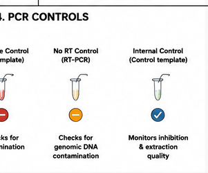
Controls are essential for interpreting molecular experiments and distinguishing genuine results from contamination or reaction failure.
Positive Control
A positive control contains a known target and should produce the expected signal when the assay is functioning properly.
Negative Control
A negative control lacks the target. It helps identify contamination or non-specific signal.
No-Template Control
In amplification assays, a no-template control contains reaction components without target nucleic acid. It is particularly useful for detecting contamination.
Internal Control
An internal control can help determine whether the sample contains inhibitory substances or whether sample processing and amplification worked appropriately.
8.26 Major Molecular Techniques at a Glance
| Technique | Main target | Major purpose |
|---|---|---|
| DNA extraction | DNA | Isolation and purification |
| RNA extraction | RNA | Isolation for expression/RNA studies |
| PCR | DNA | Target amplification |
| RT-PCR | RNA | RNA-to-cDNA conversion followed by amplification |
| qPCR | DNA/cDNA | Real-time quantitative analysis |
| Restriction digestion | DNA | Specific cleavage |
| Cloning | DNA | Propagation and study of DNA sequences |
| DNA sequencing | DNA | Nucleotide sequence determination |
| Southern blotting | DNA | Specific DNA detection |
| Northern blotting | RNA | Specific RNA detection |
| Western blotting | Protein | Specific protein detection |
| Microarray | Nucleic acids | Parallel analysis of many targets |
Important Examination Questions — Unit 8
IMPORTANT NUMERICALS AND CALCULATIONS
1. PCR Theoretical Amplification
Under ideal doubling conditions:
Example
If 100 DNA molecules are subjected to 5 ideal amplification cycles:
The theoretical number of molecules is therefore 3200.
2. Dilution Calculations in Molecular Biology
A common laboratory calculation uses:
where:
- C₁ = starting concentration
- V₁ = volume required from starting solution
- C₂ = desired concentration
- V₂ = final volume
Example
Prepare 10 mL of a 20 ng/µL DNA solution from a 100 ng/µL stock.
Therefore, 2 µL of stock is required, and the remaining volume is made up with the appropriate diluent to reach the final volume.
3. Percentage Solution Calculation
Percentage concentration may be encountered in preparation of laboratory solutions.
4. Spectrophotometric Concentration
Using Beer-Lambert law:
Therefore:
Numerical Examination Strategy
- Write the formula first.
- List the given values.
- Identify the unknown quantity.
- Check units.
- Rearrange the formula if necessary.
- Substitute values carefully.
- Show intermediate calculations.
- Write the final answer with appropriate units.
SECTION 3 — MASTER REVISION
Spectroscopy
- Spectroscopy studies interaction between radiation and matter.
- Wavelength, frequency and energy are related.
- c = λν
- E = hν
- Beer-Lambert law: A = εlc
- UV-visible spectroscopy is widely used for quantitative analysis.
- Fluorescence spectroscopy measures emitted radiation following excitation.
- Infrared spectroscopy provides information about molecular vibrations and functional groups.
Molecular Techniques
- DNA extraction isolates DNA for downstream applications.
- RNA extraction requires careful protection against RNase-mediated degradation.
- PCR amplifies selected DNA sequences.
- PCR consists of denaturation, annealing and extension.
- RT-PCR begins with RNA-to-cDNA conversion.
- qPCR monitors amplification using fluorescence.
- Restriction enzymes recognize specific DNA sequences.
- DNA sequencing determines nucleotide order.
- Southern blotting detects specific DNA sequences.
- Northern blotting detects specific RNA molecules.
- Western blotting detects specific proteins using antibodies.
- Controls are essential for reliable molecular testing.
High-Yield Long-Answer Topics
- UV-visible spectrophotometry
- Beer-Lambert law
- Fluorescence spectroscopy
- Infrared spectroscopy
- DNA extraction
- PCR
- RT-PCR
- qPCR
- Restriction enzyme digestion
- DNA cloning
- DNA sequencing
- Southern blotting
- Northern blotting
- Western blotting
- Molecular techniques in microbiological diagnosis
SECTION 3 — COMPLETE EXAM QUESTION BANK
Very Short Questions — 2 Marks
- Define spectroscopy.
- Define absorbance.
- State Beer-Lambert law.
- What is fluorescence?
- What is Stokes shift?
- Define DNA extraction.
- What is PCR?
- Name the three basic PCR steps.
- What is reverse transcription?
- Define qPCR.
- What is a restriction enzyme?
- Define DNA cloning.
- What is DNA sequencing?
- What is Southern blotting?
- What is Northern blotting?
- What is Western blotting?
- What is a positive control?
- What is a no-template control?
Short Questions — 5 Marks
- Explain electromagnetic radiation.
- Explain Beer-Lambert law.
- Describe the components of a UV-visible spectrophotometer.
- Explain the working of a spectrophotometer.
- Describe fluorescence spectroscopy.
- Explain infrared spectroscopy.
- Describe DNA extraction.
- Explain RNA extraction and its precautions.
- Explain PCR and its three basic steps.
- Describe RT-PCR.
- Explain quantitative PCR.
- Describe restriction enzyme digestion.
- Explain DNA cloning.
- Describe DNA sequencing.
- Explain Southern blotting.
- Explain Northern blotting.
- Explain Western blotting.
- Describe the importance of controls in molecular techniques.
Long Questions — 10 Marks
- Describe spectroscopy and explain the principle, instrumentation and applications of UV-visible spectrophotometry.
- Explain Beer-Lambert law in detail. Discuss its applications, limitations and numerical calculations.
- Describe fluorescence spectroscopy with principle, instrumentation, working, applications, advantages and limitations.
- Explain infrared spectroscopy and discuss its biological applications.
- Describe DNA extraction in detail, including principle, major steps, purification and applications.
- Explain PCR in detail with principle, components, cycle, amplification, applications, advantages and limitations.
- Describe RT-PCR and explain its applications in molecular biology and microbiology.
- Explain qPCR and discuss its principle, fluorescence detection and applications.
- Describe restriction enzymes and their applications in recombinant DNA technology.
- Explain DNA cloning with suitable diagram and discuss its major applications.
- Describe DNA sequencing and its importance in molecular biology.
- Explain Southern, Northern and Western blotting and compare their principles and applications.
- Discuss molecular techniques used in modern microbiological diagnosis.
Definition → Principle → Labelled diagram → Instrumentation/components → Working/mechanism → Procedure → Factors affecting the result → Applications → Advantages → Limitations → Important points → Conclusion
For numerical questions, show: Given → Formula → Substitution → Calculation → Final answer with unit.
SECTION 3 IMAGE FILE LIST
Generate or prepare the following PNG diagrams and place them inside:
| No. | PNG filename | Topic |
|---|---|---|
| 45 | 45_electromagnetic_spectrum.png | Electromagnetic spectrum |
| 46 | 46_uv_visible_spectrophotometer.png | UV-visible spectrophotometer |
| 47 | 47_fluorescence_spectroscopy.png | Fluorescence spectroscopy |
| 48 | 48_ir_spectroscopy.png | Infrared spectroscopy |
| 49 | 49_molecular_biology_workflow.png | Molecular biology workflow |
| 50 | 50_dna_extraction.png | DNA extraction |
| 51 | 51_rna_extraction.png | RNA extraction |
| 52 | 52_pcr_cycle.png | PCR cycle |
| 53 | 53_rt_pcr.png | RT-PCR |
| 54 | 54_qpcr.png | qPCR |
| 55 | 55_restriction_enzyme_digestion.png | Restriction enzyme digestion |
| 56 | 56_dna_cloning.png | DNA cloning |
| 57 | 57_dna_sequencing.png | DNA sequencing |
| 58 | 58_southern_blotting.png | Southern blotting |
| 59 | 59_northern_blotting.png | Northern blotting |
| 60 | 60_western_blotting.png | Western blotting |
| 61 | 61_dna_microarray.png | DNA microarray |
| 62 | 62_crispr_gene_editing.png | CRISPR concept |
| 63 | 63_molecular_diagnostic_workflow.png | Molecular diagnostic workflow |
| 64 | 64_pcr_controls.png | PCR controls |