UNIT 5 — ELECTROPHORETIC TECHNIQUES
5.1 Introduction to Electrophoresis
Electrophoresis is one of the most important analytical and preparative techniques used in biochemistry, molecular biology, microbiology, genetics, biotechnology and biomedical research. It is particularly useful for separating proteins, DNA, RNA and other charged biological molecules.
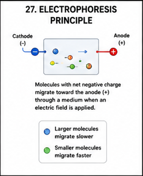
The basic principle is simple. When a charged molecule is placed in an electric field, it experiences an electrical force and moves toward the electrode carrying the opposite charge. Positively charged molecules move toward the cathode, while negatively charged molecules move toward the anode. However, the actual movement through a gel is influenced not only by electrical charge but also by molecular size and shape.
5.2 Basic Principle of Electrophoretic Migration
When a charged molecule is placed in an electric field, the electrical force acting on the molecule causes migration. The direction of movement depends on the sign of the molecular charge.
The electrophoretic mobility of a molecule is affected by the relationship between electrical driving force and resistance produced by the medium. Small molecules generally pass through a porous gel more easily than large molecules. Therefore, biological molecules can often be separated according to molecular size.
In practical electrophoresis, the migration rate is also affected by buffer pH, ionic strength, gel concentration, voltage, temperature, molecular conformation and the charge-to-mass ratio of the molecule.
5.3 Main Components of an Electrophoresis System
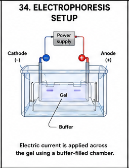
- Power supply: Provides controlled direct electrical current or voltage.
- Electrophoresis chamber: Contains the gel and running buffer.
- Electrodes: Provide the electrical field required for migration.
- Supporting medium: Usually agarose or polyacrylamide gel.
- Running buffer: Maintains appropriate pH and provides ions to conduct electricity.
- Sample loading system: Used to introduce biological samples into wells.
- Detection system: Used to visualize separated molecules.
5.4 Factors Affecting Electrophoretic Separation
- Net charge of the molecule
- Molecular size
- Molecular shape
- Buffer pH
- Buffer ionic strength
- Gel concentration
- Voltage applied
- Running time
- Temperature
- Composition of the sample
- Presence of denaturing agents
- Interaction between molecules and gel matrix
5.5 Agarose Gel Electrophoresis
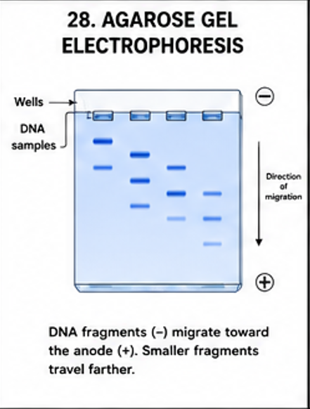
Agarose gel electrophoresis is primarily used for the separation and analysis of DNA and RNA molecules. Agarose is a polysaccharide obtained from agar and forms a porous three-dimensional network when dissolved in hot buffer and allowed to cool.
Principle
DNA molecules possess negatively charged phosphate groups. When an electric field is applied, DNA migrates toward the positive electrode. The agarose matrix acts as a molecular sieve. Smaller DNA fragments move through the pores more rapidly and therefore travel farther than larger fragments.
Preparation of Agarose Gel
- Prepare the required concentration of agarose in an appropriate electrophoresis buffer.
- Heat the mixture until the agarose dissolves completely.
- Allow the solution to cool sufficiently before adding a suitable nucleic-acid visualization reagent when appropriate.
- Place a comb in the casting tray to form wells.
- Pour the molten agarose into the casting tray.
- Allow the gel to solidify.
- Remove the comb carefully.
- Place the gel in the electrophoresis chamber.
Sample Loading
DNA samples are mixed with loading dye. The loading dye increases sample density so that the sample sinks into the wells and also provides tracking dyes that help monitor electrophoresis.
Applications of Agarose Gel Electrophoresis
- Separation of DNA fragments
- Analysis of PCR products
- Restriction enzyme analysis
- DNA fingerprinting
- Verification of cloning experiments
- Estimation of approximate DNA fragment size
- Analysis of plasmid DNA
- Analysis of RNA preparations
- Purification of selected DNA fragments
Advantages
- Simple procedure
- Relatively inexpensive
- Suitable for routine molecular biology laboratories
- Can separate a wide range of DNA fragment sizes
- Results can be visualized relatively easily
Limitations
- Resolution is lower than that of some polyacrylamide systems.
- Very small differences in fragment size may not be resolved.
- Excessive voltage may cause overheating.
- Improper gel concentration can reduce separation quality.
5.6 Polyacrylamide Gel Electrophoresis — PAGE
Polyacrylamide gel electrophoresis, commonly abbreviated as PAGE, uses a polyacrylamide matrix as the supporting medium. It provides higher resolution than agarose for many protein and small nucleic-acid applications.

Principle
Polyacrylamide forms a finely controlled porous matrix. Molecules migrate through the pores according to their charge, size and shape. The properties of the gel can be modified by changing the concentration and composition of acrylamide and cross-linking components.
Types of PAGE
- SDS-PAGE
- Native PAGE
- Isoelectric focusing
- Two-dimensional electrophoresis
5.7 SDS-PAGE
Sodium dodecyl sulfate polyacrylamide gel electrophoresis, commonly called SDS-PAGE, is one of the most widely used techniques for analyzing proteins.
Principle of SDS-PAGE
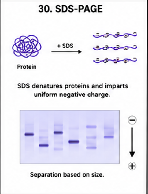
SDS is an anionic detergent that binds to hydrophobic regions of proteins and disrupts many non-covalent interactions. Together with suitable sample treatment, it causes proteins to become largely unfolded and gives them a relatively similar negative charge-to-mass relationship.
As a result, the major factor determining migration through the polyacrylamide gel becomes molecular size. Smaller proteins generally move more rapidly through the gel than larger proteins.
Common Components
- Protein sample
- SDS
- Polyacrylamide gel
- Running buffer
- Sample loading buffer
- Reducing agent when reducing conditions are required
- Protein molecular-weight marker
- Power supply
General Procedure
- Prepare protein samples.
- Mix samples with appropriate loading buffer.
- Apply heat when the experimental protocol requires denaturation.
- Load samples into gel wells.
- Load a molecular-weight marker.
- Apply an electrical field.
- Allow proteins to migrate through the gel.
- Stop electrophoresis when suitable separation has been achieved.
- Stain the gel using an appropriate protein staining method.
- Destain when required.
- Analyze the resulting protein bands.
Applications of SDS-PAGE
- Determination of approximate molecular mass of proteins
- Assessment of protein purity
- Comparison of protein expression
- Monitoring purification steps
- Analysis of recombinant proteins
- Preparation for Western blotting
- Detection of changes in protein composition
Limitations
- Protein structure is generally disrupted.
- Biological activity is not preserved under strongly denaturing conditions.
- Proteins with similar molecular masses may be difficult to distinguish.
- Post-translational modifications may complicate interpretation.
5.8 Native PAGE
Native PAGE is performed under conditions that attempt to preserve the natural structure and biological properties of proteins. Unlike SDS-PAGE, proteins are not deliberately given a uniform charge-to-mass ratio. Therefore migration depends on several properties.
These include:
- Net electrical charge
- Molecular size
- Molecular shape
- Native conformation
5.9 Isoelectric Focusing — IEF
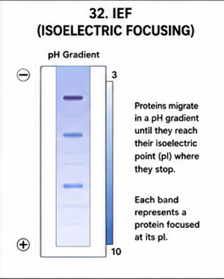
Isoelectric focusing is an electrophoretic technique used to separate proteins according to their isoelectric points.
Principle
In IEF, a stable pH gradient is established within the separation medium. A protein migrates through the gradient according to its net charge. As the protein approaches the pH corresponding to its pI, its net charge approaches zero. At this point its electrophoretic movement becomes very small and the protein focuses into a narrow band.
Importance of IEF
- Separation of proteins with different pI values
- High-resolution protein analysis
- Protein characterization
- First dimension of two-dimensional electrophoresis
- Detection of protein isoforms
5.10 Two-Dimensional Gel Electrophoresis
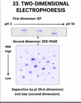
Two-dimensional electrophoresis, commonly called 2D electrophoresis, combines two different separation principles. It is especially useful for resolving complex mixtures containing many proteins.
First Dimension
Proteins are separated by isoelectric focusing according to their isoelectric points.
Second Dimension
The focused proteins are transferred to an SDS-polyacrylamide gel and separated mainly according to molecular mass.
Applications
- Proteomics
- Comparison of protein expression patterns
- Identification of protein isoforms
- Disease-associated protein studies
- Analysis of complex biological samples
- Comparative analysis of treated and untreated cells
Advantages
- Very high resolving power
- Separates proteins according to two independent properties
- Useful for complex protein mixtures
- Suitable for proteomic studies
Limitations
- Technically more complicated
- Requires careful sample preparation
- Some proteins are poorly represented
- Membrane proteins can be difficult to analyze
- Quantitative interpretation can be challenging
5.11 General Electrophoresis Setup
5.12 Comparison of Major Electrophoretic Techniques
| Technique | Major molecule | Main basis of separation | Important use |
|---|---|---|---|
| Agarose electrophoresis | DNA/RNA | Mainly size | DNA fragment analysis |
| SDS-PAGE | Proteins | Mainly molecular mass | Protein size and purity |
| Native PAGE | Proteins | Charge, size and shape | Native protein analysis |
| IEF | Proteins | Isoelectric point | Protein pI analysis |
| 2D electrophoresis | Proteins | pI followed by molecular mass | Proteomics |
Important Examination Questions — Unit 5
UNIT 6 — CHROMATOGRAPHIC TECHNIQUES
6.1 Introduction to Chromatography
Chromatography is one of the most versatile techniques used in biological research. It can be used to separate, identify, purify and sometimes quantify components of complex biological mixtures.
Proteins, peptides, amino acids, nucleotides, carbohydrates, lipids, pigments, drugs, metabolites and many other compounds can be analyzed using appropriate chromatographic methods.
6.2 Basic Principle
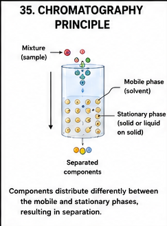
Chromatographic separation occurs because different molecules interact differently with the stationary phase and mobile phase. A molecule that has a stronger interaction with the stationary phase generally moves more slowly, while a molecule that remains more strongly associated with the mobile phase moves more rapidly.
6.3 Important Components
- Stationary phase: The phase that remains fixed.
- Mobile phase: The phase that moves through or over the stationary phase.
- Sample: Mixture containing compounds to be separated.
- Detector: Detects components as they emerge or are visualized.
- Collection system: Collects separated fractions when preparative separation is performed.
6.4 Classification of Chromatography
- Paper chromatography
- Thin-layer chromatography
- Column chromatography
- Ion-exchange chromatography
- Size-exclusion chromatography
- Affinity chromatography
- High-performance liquid chromatography
- Gas chromatography
6.5 Paper Chromatography
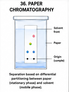
Paper chromatography is a simple chromatographic method historically used for separating small molecules such as amino acids, sugars and pigments.
Principle
The sample components distribute differently between the stationary environment associated with the paper and the moving solvent. Different components therefore migrate different distances.
Rf Value
The relative mobility of a component can be expressed using the retardation factor, commonly written as Rf.
Important Points About Rf
- Rf generally lies between 0 and 1.
- It depends on solvent composition.
- It depends on the stationary phase.
- It depends on temperature and experimental conditions.
- It can help compare an unknown compound with standards under identical conditions.
Applications
- Amino acid identification
- Pigment separation
- Carbohydrate analysis
- Teaching laboratory demonstrations
- Preliminary qualitative analysis
6.6 Thin-Layer Chromatography — TLC
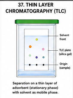
Thin-layer chromatography is a simple and rapid technique in which the stationary phase is coated as a thin layer over a solid support. Silica gel and alumina are commonly used stationary phases.
General Procedure
- Prepare or obtain the TLC plate.
- Draw a light baseline near the lower edge.
- Apply a small amount of sample.
- Place the plate in the developing chamber.
- Allow the solvent to move upward.
- Remove the plate before the solvent reaches the top.
- Mark the solvent front.
- Visualize separated compounds.
- Calculate Rf values where appropriate.
Applications
- Monitoring chemical reactions
- Analysis of amino acids and metabolites
- Drug analysis
- Pigment separation
- Checking purity of compounds
- Identification by comparison with standards
6.7 Column Chromatography
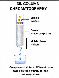
Column chromatography is a technique in which the stationary phase is packed into a column and the mobile phase passes through it. Components of a mixture move at different rates and are collected in separate fractions.
General Steps
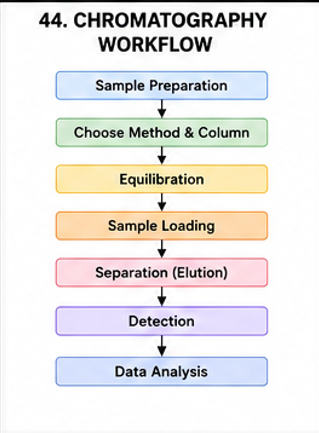
- Prepare and pack the column.
- Equilibrate the stationary phase.
- Apply the sample carefully.
- Add mobile phase.
- Allow components to migrate.
- Collect fractions.
- Analyze the fractions.
- Pool desired fractions when appropriate.
Applications
- Protein purification
- Pigment separation
- Purification of metabolites
- Isolation of chemical compounds
- Preparative separation
6.8 Ion-Exchange Chromatography
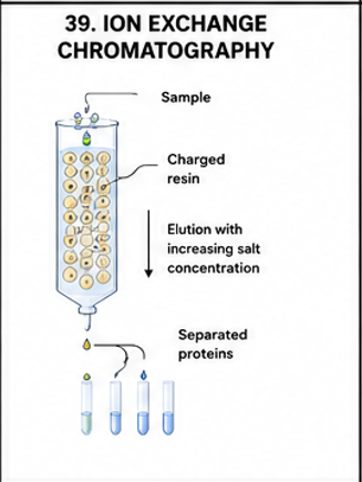
Ion-exchange chromatography separates molecules according to their electrical charge. The stationary phase contains charged functional groups that interact with oppositely charged molecules.
Types
- Cation-exchange chromatography: The stationary phase carries negative groups and binds positively charged molecules.
- Anion-exchange chromatography: The stationary phase carries positive groups and binds negatively charged molecules.
Elution
Bound molecules may be released by changing the salt concentration, changing pH or modifying other conditions that alter electrostatic interactions.
Applications
- Protein purification
- Separation of amino acids
- Purification of nucleotides
- Separation of charged metabolites
- Biopharmaceutical purification
6.9 Size-Exclusion Chromatography
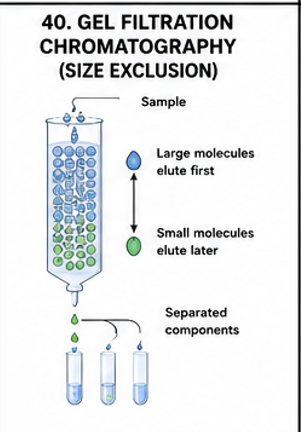
Size-exclusion chromatography separates molecules primarily according to their hydrodynamic size. It is also known as gel filtration when aqueous systems are used.
Principle
The stationary phase contains porous beads. Large molecules cannot enter many of the pores and therefore travel through the column more directly. Smaller molecules can enter more pores and consequently take a longer effective path through the column.
Applications
- Protein purification
- Protein molecular-size estimation
- Removal of small molecules
- Buffer exchange
- Separation of protein aggregates
6.10 Affinity Chromatography
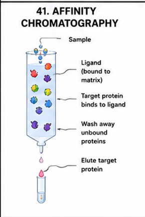
Affinity chromatography is a highly selective purification technique based on a specific biological interaction between a target molecule and a ligand attached to the stationary phase.
Principle
The target molecule specifically recognizes and binds to an immobilized ligand. Other molecules that do not bind specifically pass through the column. The target is subsequently eluted by changing appropriate conditions.
Examples
- Antigen-antibody interaction
- Enzyme-substrate or enzyme-inhibitor interaction
- Receptor-ligand interaction
- His-tagged protein and immobilized metal interaction
- Biotin-streptavidin interaction
Advantages
- Very high selectivity
- Can produce highly enriched target proteins
- Useful for recombinant protein purification
- Often reduces the number of purification steps
Limitations
- Ligands can be expensive.
- Conditions must preserve the biological interaction.
- Elution conditions may affect sensitive proteins.
- Column preparation can require optimization.
6.11 High-Performance Liquid Chromatography — HPLC
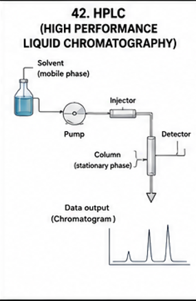
High-performance liquid chromatography is an advanced chromatographic technique in which a liquid mobile phase is forced through a packed stationary phase under relatively high pressure.
Main Components of HPLC
- Solvent reservoirs
- Degassing system
- High-pressure pump
- Sample injector
- Chromatographic column
- Detector
- Data acquisition system
Working
The mobile phase is delivered under pressure through the chromatographic column. The sample is introduced into the mobile phase and passes through the stationary phase. Components interact differently with the column and therefore elute at different times.
Applications of HPLC
- Drug analysis
- Metabolite analysis
- Protein and peptide analysis
- Food analysis
- Pharmaceutical quality control
- Biochemical research
- Clinical laboratory analysis
6.12 Gas Chromatography — GC
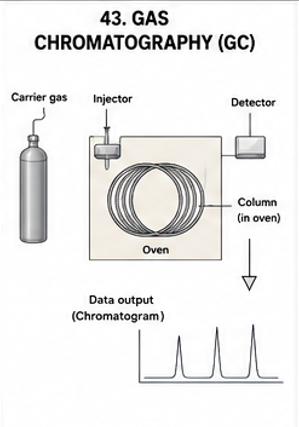
Gas chromatography is a chromatographic technique used primarily for compounds that can be vaporized without decomposition or that can be converted into suitable volatile derivatives.
Major Components
- Carrier gas supply
- Injector
- Column
- Oven
- Detector
- Data recording system
Applications
- Analysis of volatile compounds
- Metabolite analysis
- Environmental analysis
- Food analysis
- Drug analysis
- Forensic analysis
6.13 General Chromatographic Workflow
6.14 Comparison of Important Chromatographic Techniques
| Technique | Major basis | Common application |
|---|---|---|
| Paper chromatography | Differential partition/interaction | Amino acids, pigments |
| TLC | Adsorption/partition | Small molecules and reaction monitoring |
| Column chromatography | Differential interaction with stationary phase | Preparative purification |
| Ion exchange | Charge | Protein purification |
| Size exclusion | Hydrodynamic size | Protein separation and desalting |
| Affinity | Specific biological interaction | Highly selective protein purification |
| HPLC | Different retention in high-pressure liquid system | Analytical and preparative applications |
| GC | Volatility and interaction with stationary phase | Volatile compounds |
Important Examination Questions — Unit 6
IMPORTANT CALCULATIONS AND RESULT INTERPRETATION
1. Calculation of Rf Value
A common numerical question in chromatography involves calculation of the Rf value.
Example 1
Suppose a compound travels 4.0 cm from the origin while the solvent front travels 8.0 cm.
Therefore, the Rf value of the compound is 0.50.
Example 2
A compound travels 3.5 cm while the solvent front travels 7.0 cm.
Therefore, the Rf value is 0.50.
Example 3
An unknown sample has travelled 5.2 cm and the solvent front has travelled 8.0 cm.
The Rf value is therefore 0.65.
Calculation Tips for Rf Numericals
- Always measure distance from the original spotting line.
- Measure the solvent front separately.
- Do not use the total length of the TLC plate.
- Rf has no physical unit.
- Under identical experimental conditions, Rf values can help compare unknown and standard compounds.
- Rf should normally be between 0 and 1.
2. Interpretation of Electrophoresis Bands
When interpreting an electrophoresis gel, the position, intensity and number of bands should be considered.
- Band position: Gives information about relative migration and, depending on the technique, molecular size or charge.
- Band intensity: Can provide qualitative or semi-quantitative information about the amount of material, although intensity must be interpreted carefully.
- Number of bands: May indicate the number of detectable components.
- Smearing: May indicate degraded samples, overloading, inappropriate running conditions or other technical issues.
3. Approximate Protein Molecular Mass from SDS-PAGE
In SDS-PAGE, the migration of a protein can be compared with molecular mass standards. A calibration plot can be prepared using the migration distance or relative mobility of known standards.
A common approach is to plot the logarithm of molecular mass against relative migration and use the resulting calibration relationship to estimate the molecular mass of an unknown protein.
4. Chromatographic Elution Profile
In instrumental chromatography, separated compounds may appear as peaks on a chromatogram. Each peak represents a detected component or group of components under the experimental conditions.
Important features include:
- Retention time
- Peak height
- Peak area
- Peak width
- Resolution between adjacent peaks
5. Retention Time
Retention time can be useful for identifying compounds by comparison with standards analyzed under the same conditions. Identification should not normally be based on retention time alone when definitive identification is required.
MASTER COMPARISON — ELECTROPHORESIS VS CHROMATOGRAPHY
| Feature | Electrophoresis | Chromatography |
|---|---|---|
| Basic principle | Migration in an electric field | Differential distribution between stationary and mobile phases |
| Major driving factor | Electrical field | Movement of mobile phase |
| Common molecules | DNA, RNA, proteins | Proteins, metabolites, drugs, pigments and many other compounds |
| Common supporting system | Agarose or polyacrylamide gel | Column, plate, paper or specialized chromatographic column |
| Separation basis | Charge, size, shape or pI depending on method | Charge, size, affinity, adsorption, partition or volatility |
| Major application | Analysis of nucleic acids and proteins | Separation, purification and analysis of many biological compounds |
SECTION 2 — QUICK REVISION POINTS
Electrophoresis
- Electrophoresis separates charged molecules using an electric field.
- DNA is negatively charged because of its phosphate backbone.
- Agarose is commonly used for DNA and RNA separation.
- PAGE provides high-resolution separation.
- SDS-PAGE mainly separates proteins according to molecular mass.
- Native PAGE preserves more of the native properties of proteins.
- IEF separates proteins according to isoelectric point.
- 2D electrophoresis combines IEF and SDS-PAGE.
Chromatography
- Chromatography uses stationary and mobile phases.
- Different molecules interact differently with the two phases.
- Rf is important in planar chromatography.
- Ion-exchange chromatography separates according to charge.
- Size-exclusion chromatography separates according to hydrodynamic size.
- Affinity chromatography is based on specific biological interactions.
- HPLC uses high-pressure liquid flow through a chromatographic column.
- GC is particularly useful for volatile compounds.
High-Yield Long-Answer Topics
- Principle, instrumentation and applications of electrophoresis.
- Agarose gel electrophoresis.
- SDS-PAGE.
- Native PAGE and its comparison with SDS-PAGE.
- Isoelectric focusing.
- Two-dimensional electrophoresis.
- General principles of chromatography.
- Ion-exchange chromatography.
- Size-exclusion chromatography.
- Affinity chromatography.
- HPLC.
- Comparison of chromatographic techniques.
SECTION 2 — EXAM-ORIENTED QUESTION BANK
Very Short Questions — 2 Marks
- Define electrophoresis.
- What is electrophoretic mobility?
- What is agarose?
- Define SDS-PAGE.
- What is Native PAGE?
- Define isoelectric point.
- What is isoelectric focusing?
- What is two-dimensional electrophoresis?
- Define chromatography.
- What is stationary phase?
- What is mobile phase?
- Define Rf value.
- What is ion-exchange chromatography?
- What is gel filtration?
- Define affinity chromatography.
- What is HPLC?
- What is gas chromatography?
- Define retention time.
Short Questions — 5 Marks
- Explain the principle of agarose gel electrophoresis.
- Describe the basic components of an electrophoresis apparatus.
- Explain SDS-PAGE.
- Differentiate Native PAGE and SDS-PAGE.
- Explain isoelectric focusing.
- Describe two-dimensional electrophoresis.
- Explain paper chromatography.
- Describe thin-layer chromatography.
- Explain ion-exchange chromatography.
- Describe size-exclusion chromatography.
- Explain affinity chromatography.
- Describe the major components of HPLC.
- Explain gas chromatography.
Long Questions — 10 Marks
- Describe electrophoresis in detail, including its principle, instrumentation, factors affecting migration, major types and applications.
- Explain agarose gel electrophoresis with principle, gel preparation, sample loading, working, applications, advantages and limitations.
- Describe SDS-PAGE in detail and explain how proteins are separated according to molecular mass.
- Explain Native PAGE and compare it with SDS-PAGE.
- Describe isoelectric focusing and explain its principle and applications.
- Explain two-dimensional gel electrophoresis with a suitable diagram and discuss its importance in proteomics.
- Define chromatography and describe its general principle, components and classification.
- Explain ion-exchange chromatography in detail with principle, types, working and applications.
- Describe size-exclusion chromatography with principle, working and applications.
- Explain affinity chromatography in detail with principle, working, applications, advantages and limitations.
- Describe HPLC with principle, instrumentation, working and applications.
- Discuss important chromatographic techniques used in biological research.